• 文献检索
  • 文档翻译
  • 深度研究
  • 学术资讯
  • Suppr Zotero 插件Zotero 插件
  • 邀请有礼
  • 套餐&价格
  • 历史记录
应用&插件
Suppr Zotero 插件Zotero 插件浏览器插件Mac 客户端Windows 客户端微信小程序
定价
高级版会员购买积分包购买API积分包
服务
文献检索文档翻译深度研究API 文档MCP 服务
关于我们
关于 Suppr公司介绍联系我们用户协议隐私条款
关注我们

Suppr 超能文献

核心技术专利:CN118964589B侵权必究
粤ICP备2023148730 号-1Suppr @ 2026

文献检索

告别复杂PubMed语法,用中文像聊天一样搜索,搜遍4000万医学文献。AI智能推荐,让科研检索更轻松。

立即免费搜索

文件翻译

保留排版,准确专业,支持PDF/Word/PPT等文件格式,支持 12+语言互译。

免费翻译文档

深度研究

AI帮你快速写综述,25分钟生成高质量综述,智能提取关键信息,辅助科研写作。

立即免费体验

树突状细胞(pDCs)对 SARS-CoV-2 的感应及其随后产生的 IFN-I,有助于 COVID-19 期间巨噬细胞诱导的细胞因子风暴。

Sensing of SARS-CoV-2 by pDCs and their subsequent production of IFN-I contribute to macrophage-induced cytokine storm during COVID-19.

机构信息

HSS Research Institute and David Z. Rosensweig Genomics Research Center, Hospital for Special Surgery, New York, NY 10021, USA.

Institute for Computational Biomedicine and Caryl and Israel Englander Institute for Precision Medicine, Weill Cornell Medicine, New York, NY 10021, USA.

出版信息

Sci Immunol. 2022 Sep 9;7(75):eadd4906. doi: 10.1126/sciimmunol.add4906.

DOI:10.1126/sciimmunol.add4906
PMID:36083891
原文链接:https://pmc.ncbi.nlm.nih.gov/articles/PMC9853436/
Abstract

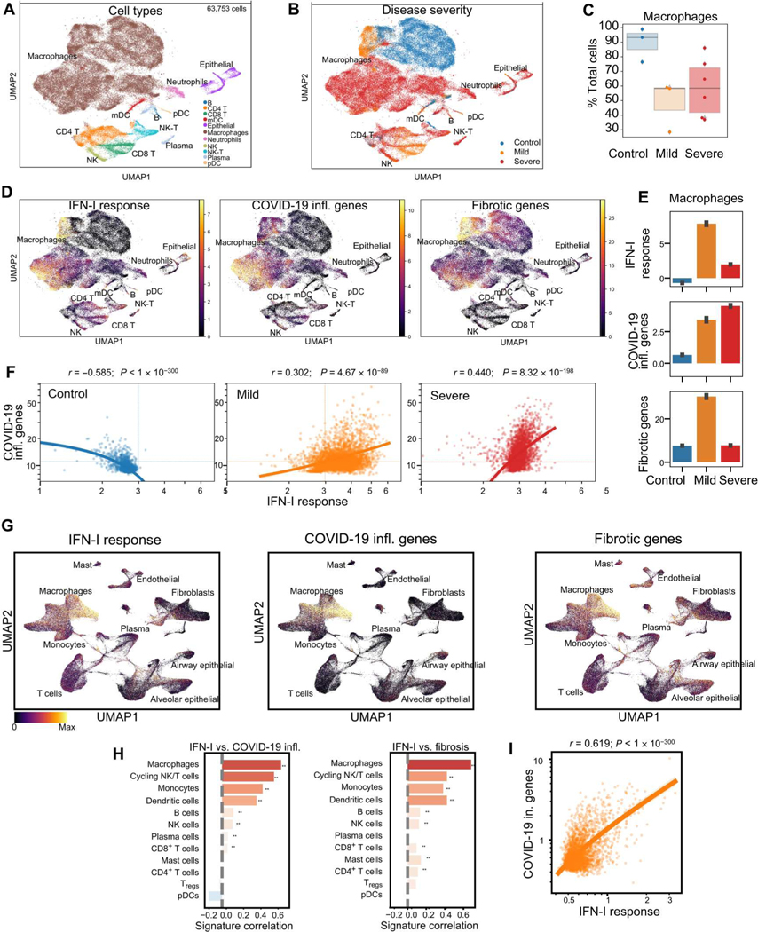
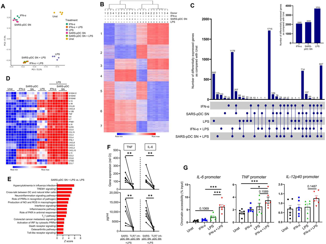
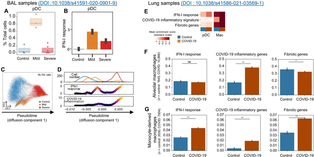
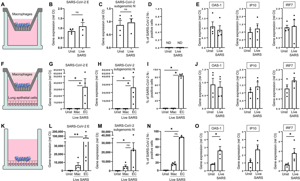
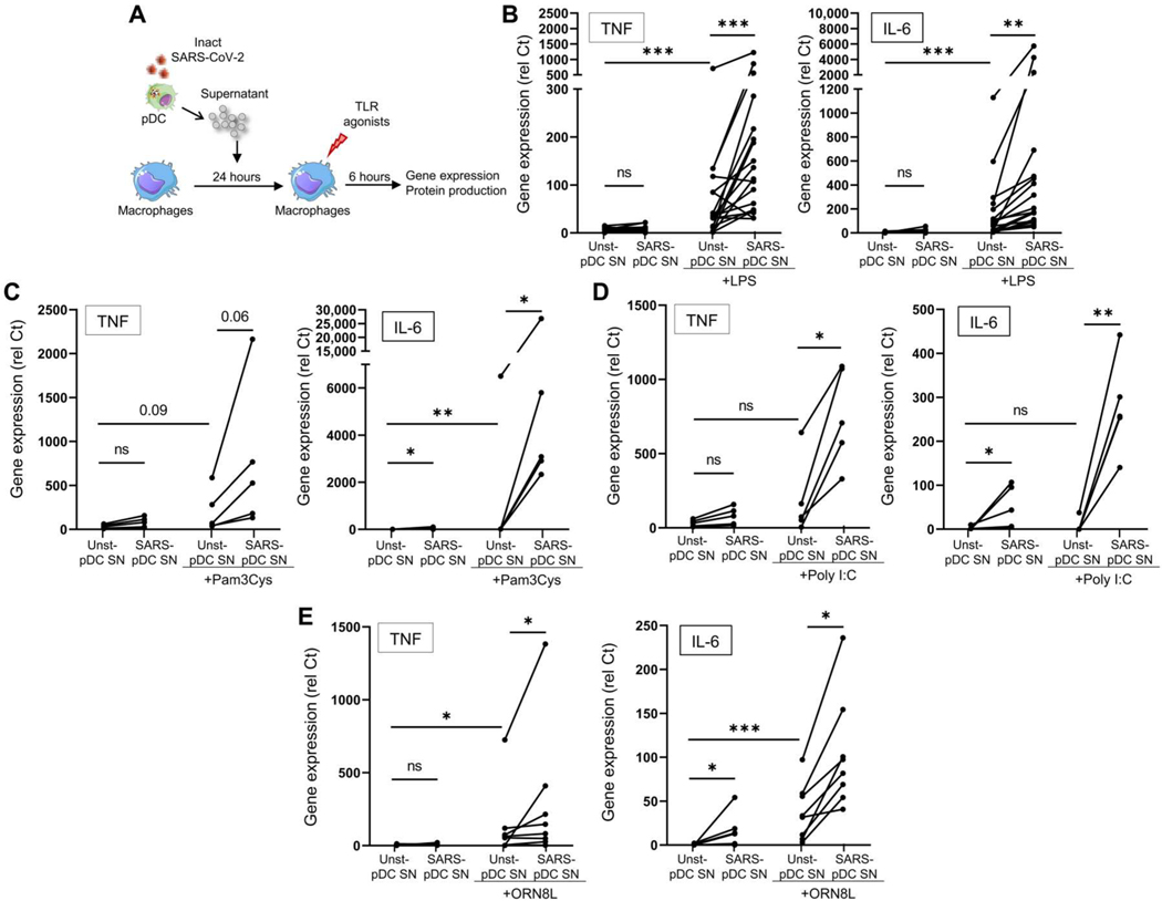
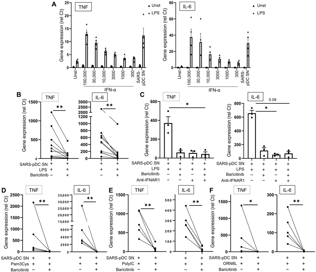
Lung-infiltrating macrophages create a marked inflammatory milieu in a subset of patients with COVID-19 by producing a cytokine storm, which correlates with increased lethality. However, these macrophages are largely not infected by SARS-CoV-2, so the mechanism underlying their activation in the lung is unclear. Type I interferons (IFN-I) contribute to protecting the host against SARS-CoV-2 but may also have some deleterious effect, and the source of IFN-I in the lungs of infected patients is not well defined. Plasmacytoid dendritic cells (pDCs), a key cell type involved in antiviral responses, can produce IFN-I in response to SARS-CoV-2. We observed the infiltration of pDCs in the lungs of SARS-CoV-2-infected patients, which correlated with strong IFN-I signaling in lung macrophages. In patients with severe COVID-19, lung macrophages expressed a robust inflammatory signature, which correlated with persistent IFN-I signaling at the single-cell level. Hence, we observed the uncoupling in the kinetics of the infiltration of pDCs in the lungs and the associated IFN-I signature, with the cytokine storm in macrophages. We observed that pDCs were the dominant IFN-α-producing cells in response to the virus in the blood, whereas macrophages produced IFN-α only when in physical contact with infected epithelial cells. We also showed that IFN-α produced by pDCs, after the sensing of SARS-CoV-2 by TLR7, mediated changes in macrophages at both transcriptional and epigenetic levels, which favored their hyperactivation by environmental stimuli. Together, these data indicate that the priming of macrophages can result from the response by pDCs to SARS-CoV-2, leading to macrophage activation in patients with severe COVID-19.

摘要

肺浸润性巨噬细胞通过产生细胞因子风暴在 COVID-19 的一部分患者中造成明显的炎症环境,这与致死率增加相关。然而,这些巨噬细胞在很大程度上不受 SARS-CoV-2 的感染,因此它们在肺部中的激活机制尚不清楚。I 型干扰素(IFN-I)有助于宿主抵抗 SARS-CoV-2,但也可能产生一些有害影响,而感染患者肺部中 IFN-I 的来源尚未明确。浆细胞样树突状细胞(pDCs)是参与抗病毒反应的关键细胞类型,可对 SARS-CoV-2 产生 IFN-I。我们观察到 SARS-CoV-2 感染患者的肺中 pDCs 的浸润,这与肺巨噬细胞中强烈的 IFN-I 信号相关。在 COVID-19 重症患者中,肺巨噬细胞表达强烈的炎症特征,这与单细胞水平上持续的 IFN-I 信号相关。因此,我们观察到 pDCs 在肺部的浸润和相关 IFN-I 特征与巨噬细胞中的细胞因子风暴之间的动力学失耦。我们观察到 pDCs 是血液中针对病毒产生 IFN-α的主要细胞,而巨噬细胞仅在与受感染的上皮细胞物理接触时才产生 IFN-α。我们还表明,pDCs 在 TLR7 感知 SARS-CoV-2 后产生的 IFN-α在转录和表观遗传水平上改变了巨噬细胞,这有利于它们被环境刺激过度激活。总之,这些数据表明,巨噬细胞的激活可以源于 pDCs 对 SARS-CoV-2 的反应,导致 COVID-19 重症患者的巨噬细胞激活。

https://cdn.ncbi.nlm.nih.gov/pmc/blobs/b938/9853436/21030f97d016/nihms-1843099-f0008.jpg
https://cdn.ncbi.nlm.nih.gov/pmc/blobs/b938/9853436/e5a32193356b/nihms-1843099-f0001.jpg
https://cdn.ncbi.nlm.nih.gov/pmc/blobs/b938/9853436/cb4ba7ca6b4a/nihms-1843099-f0002.jpg
https://cdn.ncbi.nlm.nih.gov/pmc/blobs/b938/9853436/c173c3d7056c/nihms-1843099-f0003.jpg
https://cdn.ncbi.nlm.nih.gov/pmc/blobs/b938/9853436/0690d24885e2/nihms-1843099-f0004.jpg
https://cdn.ncbi.nlm.nih.gov/pmc/blobs/b938/9853436/e62613654181/nihms-1843099-f0005.jpg
https://cdn.ncbi.nlm.nih.gov/pmc/blobs/b938/9853436/47b0d845a11b/nihms-1843099-f0006.jpg
https://cdn.ncbi.nlm.nih.gov/pmc/blobs/b938/9853436/76306483afff/nihms-1843099-f0007.jpg
https://cdn.ncbi.nlm.nih.gov/pmc/blobs/b938/9853436/21030f97d016/nihms-1843099-f0008.jpg

相似文献

1
Sensing of SARS-CoV-2 by pDCs and their subsequent production of IFN-I contribute to macrophage-induced cytokine storm during COVID-19.树突状细胞(pDCs)对 SARS-CoV-2 的感应及其随后产生的 IFN-I,有助于 COVID-19 期间巨噬细胞诱导的细胞因子风暴。
Sci Immunol. 2022 Sep 9;7(75):eadd4906. doi: 10.1126/sciimmunol.add4906.
2
Macrophage phagocytosis of SARS-CoV-2-infected cells mediates potent plasmacytoid dendritic cell activation.巨噬细胞吞噬 SARS-CoV-2 感染的细胞可介导强烈的浆细胞样树突状细胞激活。
Cell Mol Immunol. 2023 Jul;20(7):835-849. doi: 10.1038/s41423-023-01039-4. Epub 2023 May 30.
3
Plasmacytoid dendritic cells produce type I interferon and reduce viral replication in airway epithelial cells after SARS-CoV-2 infection.浆细胞样树突状细胞在感染新型冠状病毒后产生I型干扰素并减少气道上皮细胞中的病毒复制。
bioRxiv. 2021 May 13:2021.05.12.443948. doi: 10.1101/2021.05.12.443948.
4
High secretion of interferons by human plasmacytoid dendritic cells upon recognition of Middle East respiratory syndrome coronavirus.人浆细胞样树突状细胞识别中东呼吸综合征冠状病毒后干扰素分泌增加
J Virol. 2015 Apr;89(7):3859-69. doi: 10.1128/JVI.03607-14. Epub 2015 Jan 21.
5
Severe COVID-19 patients have impaired plasmacytoid dendritic cell-mediated control of SARS-CoV-2.重症 COVID-19 患者的浆细胞样树突状细胞介导的 SARS-CoV-2 控制受损。
Nat Commun. 2023 Feb 8;14(1):694. doi: 10.1038/s41467-023-36140-9.
6
Differential plasmacytoid dendritic cell phenotype and type I Interferon response in asymptomatic and severe COVID-19 infection.无症状和重症 COVID-19 感染中的浆细胞样树突状细胞表型和 I 型干扰素反应的差异。
PLoS Pathog. 2021 Sep 2;17(9):e1009878. doi: 10.1371/journal.ppat.1009878. eCollection 2021 Sep.
7
TLR2 and TLR7 mediate distinct immunopathological and antiviral plasmacytoid dendritic cell responses to SARS-CoV-2 infection.Toll样受体2(TLR2)和Toll样受体7(TLR7)介导浆细胞样树突状细胞对严重急性呼吸综合征冠状病毒2(SARS-CoV-2)感染产生不同的免疫病理反应和抗病毒反应。
EMBO J. 2022 May 16;41(10):e109622. doi: 10.15252/embj.2021109622. Epub 2022 Mar 1.
8
Janus kinase signaling as risk factor and therapeutic target for severe SARS-CoV-2 infection.Janus 激酶信号作为严重 SARS-CoV-2 感染的风险因素和治疗靶点。
Eur J Immunol. 2021 May;51(5):1071-1075. doi: 10.1002/eji.202149173. Epub 2021 Mar 22.
9
A Network-Based Analysis Reveals the Mechanism Underlying Vitamin D in Suppressing Cytokine Storm and Virus in SARS-CoV-2 Infection.基于网络的分析揭示了维生素 D 抑制 SARS-CoV-2 感染中细胞因子风暴和病毒的机制。
Front Immunol. 2020 Dec 9;11:590459. doi: 10.3389/fimmu.2020.590459. eCollection 2020.
10
Plasmacytoid dendritic cells during COVID-19: Ally or adversary?COVID-19 期间的浆细胞样树突状细胞:盟友还是敌人?
Cell Rep. 2022 Jul 26;40(4):111148. doi: 10.1016/j.celrep.2022.111148. Epub 2022 Jul 14.

引用本文的文献

1
Glutamate utilization fuels rapid production of mitochondrial ROS in dendritic cells and drives systemic inflammation during tularemia.谷氨酸的利用为树突状细胞中线粒体活性氧的快速产生提供能量,并在兔热病期间引发全身炎症。
Sci Adv. 2025 Aug 29;11(35):eadu6271. doi: 10.1126/sciadv.adu6271.
2
Artificial liver plasma adsorption improves survival in elderly patients with severe pneumonia: a retrospective cohort study.人工肝血浆吸附改善老年重症肺炎患者的生存率:一项回顾性队列研究。
Front Med (Lausanne). 2025 Jul 9;12:1613810. doi: 10.3389/fmed.2025.1613810. eCollection 2025.
3
Phagocytic activity of blood monocytes and neutrophils in moderate COVID-19 patients and impact of immune therapy with bacterial lysates.

本文引用的文献

1
CXCL4 synergizes with TLR8 for TBK1-IRF5 activation, epigenomic remodeling and inflammatory response in human monocytes.CXCL4 与 TLR8 协同作用,激活人类单核细胞中的 TBK1-IRF5,重塑表观基因组并引发炎症反应。
Nat Commun. 2022 Jun 14;13(1):3426. doi: 10.1038/s41467-022-31132-7.
2
Chemokines form nanoparticles with DNA and can superinduce TLR-driven immune inflammation.趋化因子与 DNA 形成纳米颗粒,并能超诱导 TLR 驱动的免疫炎症。
J Exp Med. 2022 Jul 4;219(7). doi: 10.1084/jem.20212142. Epub 2022 May 31.
3
ZBP1-dependent inflammatory cell death, PANoptosis, and cytokine storm disrupt IFN therapeutic efficacy during coronavirus infection.
中度新冠肺炎患者血液单核细胞和中性粒细胞的吞噬活性以及细菌裂解物免疫疗法的影响
PLoS One. 2025 Jun 5;20(6):e0324987. doi: 10.1371/journal.pone.0324987. eCollection 2025.
4
TLR7 deficiency enhances inflammation in the URT but reduces LRT immunity following influenza A infection.Toll样受体7(TLR7)缺陷增强了上呼吸道(URT)的炎症反应,但降低了甲型流感病毒感染后下呼吸道(LRT)的免疫力。
Sci Rep. 2025 May 29;15(1):18918. doi: 10.1038/s41598-025-04154-6.
5
Interferon-β treatment reverses the detrimental effect of B-cell depletion therapy on respiratory virus infection.干扰素-β治疗可逆转B细胞清除疗法对呼吸道病毒感染的有害影响。
J Immunol. 2025 Jul 1;214(7):1688-1697. doi: 10.1093/jimmun/vkaf085.
6
Enhanced TLR7-dependent production of type I interferon by pDCs underlies pandemic chilblains.浆细胞样树突状细胞(pDCs)中由Toll样受体7(TLR7)依赖性增强的I型干扰素产生是大流行冻疮的基础。
J Exp Med. 2025 Jul 7;222(7). doi: 10.1084/jem.20231467. Epub 2025 Apr 14.
7
Toll-like receptor 7 (TLR7)-mediated antiviral response protects mice from lethal SARS-CoV-2 infection.Toll样受体7(TLR7)介导的抗病毒反应可保护小鼠免受致命的严重急性呼吸综合征冠状病毒2(SARS-CoV-2)感染。
J Virol. 2025 May 20;99(5):e0166824. doi: 10.1128/jvi.01668-24. Epub 2025 Mar 31.
8
Exploring the Contribution of TLR7 to Sex-Based Disparities in Respiratory Syncytial Virus (RSV)-Induced Inflammation and Immunity.探索Toll样受体7(TLR7)在呼吸道合胞病毒(RSV)诱导的炎症和免疫中基于性别的差异所起的作用。
Viruses. 2025 Mar 16;17(3):428. doi: 10.3390/v17030428.
9
Cytosolic nucleic acid sensing as driver of critical illness: mechanisms and advances in therapy.胞质核酸感应作为危重症的驱动因素:机制与治疗进展
Signal Transduct Target Ther. 2025 Mar 19;10(1):90. doi: 10.1038/s41392-025-02174-2.
10
Immunologic and inflammatory consequences of SARS-CoV-2 infection and its implications in renal disease.严重急性呼吸综合征冠状病毒2(SARS-CoV-2)感染的免疫和炎症后果及其在肾脏疾病中的意义。
Front Immunol. 2025 Feb 12;15:1376654. doi: 10.3389/fimmu.2024.1376654. eCollection 2024.
ZBP1 依赖性炎症细胞死亡、PANoptosis 和细胞因子风暴破坏了冠状病毒感染期间 IFN 治疗的疗效。
Sci Immunol. 2022 Aug 26;7(74):eabo6294. doi: 10.1126/sciimmunol.abo6294.
4
Inflammasome activation in infected macrophages drives COVID-19 pathology.在被感染的巨噬细胞中激活炎症小体导致 COVID-19 病理学。
Nature. 2022 Jun;606(7914):585-593. doi: 10.1038/s41586-022-04802-1. Epub 2022 Apr 28.
5
Inflammatory responses in the placenta upon SARS-CoV-2 infection late in pregnancy.妊娠晚期感染新冠病毒后胎盘的炎症反应。
iScience. 2022 May 20;25(5):104223. doi: 10.1016/j.isci.2022.104223. Epub 2022 Apr 11.
6
The immunology and immunopathology of COVID-19.新型冠状病毒肺炎的免疫学和免疫病理学。
Science. 2022 Mar 11;375(6585):1122-1127. doi: 10.1126/science.abm8108. Epub 2022 Mar 10.
7
TLR2 and TLR7 mediate distinct immunopathological and antiviral plasmacytoid dendritic cell responses to SARS-CoV-2 infection.Toll样受体2(TLR2)和Toll样受体7(TLR7)介导浆细胞样树突状细胞对严重急性呼吸综合征冠状病毒2(SARS-CoV-2)感染产生不同的免疫病理反应和抗病毒反应。
EMBO J. 2022 May 16;41(10):e109622. doi: 10.15252/embj.2021109622. Epub 2022 Mar 1.
8
Interferon-γ primes macrophages for pathogen ligand-induced killing via a caspase-8 and mitochondrial cell death pathway.干扰素-γ 通过半胱天冬酶-8 和线粒体细胞死亡途径诱导巨噬细胞对病原体配体的杀伤作用。
Immunity. 2022 Mar 8;55(3):423-441.e9. doi: 10.1016/j.immuni.2022.01.003. Epub 2022 Feb 8.
9
Extensive mucosal sloughing of the small intestine and colon in a patient with severe COVID-19.一名重症新型冠状病毒肺炎患者出现广泛的小肠和结肠黏膜脱落。
DEN Open. 2022 Apr;2(1):e42. doi: 10.1002/deo2.42. Epub 2021 Sep 7.
10
The cGAS-STING pathway drives type I IFN immunopathology in COVID-19.cGAS-STING 通路驱动 COVID-19 中的 I 型 IFN 免疫病理学。
Nature. 2022 Mar;603(7899):145-151. doi: 10.1038/s41586-022-04421-w. Epub 2022 Jan 19.