Department of Pediatrics, Emory University School of Medicine and Children's Healthcare of Atlanta, Atlanta, GA, USA.
Techshot, Inc., Greenville, IN, USA.
Stem Cell Reports. 2022 Oct 11;17(10):2272-2285. doi: 10.1016/j.stemcr.2022.08.007. Epub 2022 Sep 8.
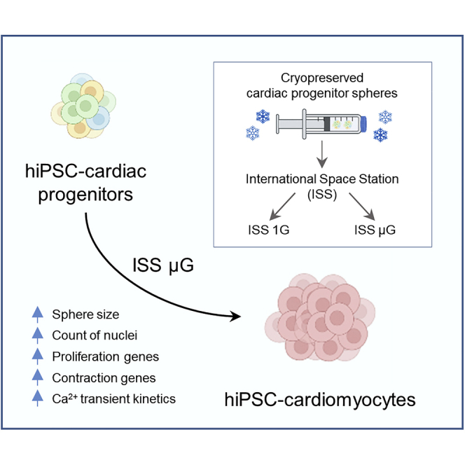
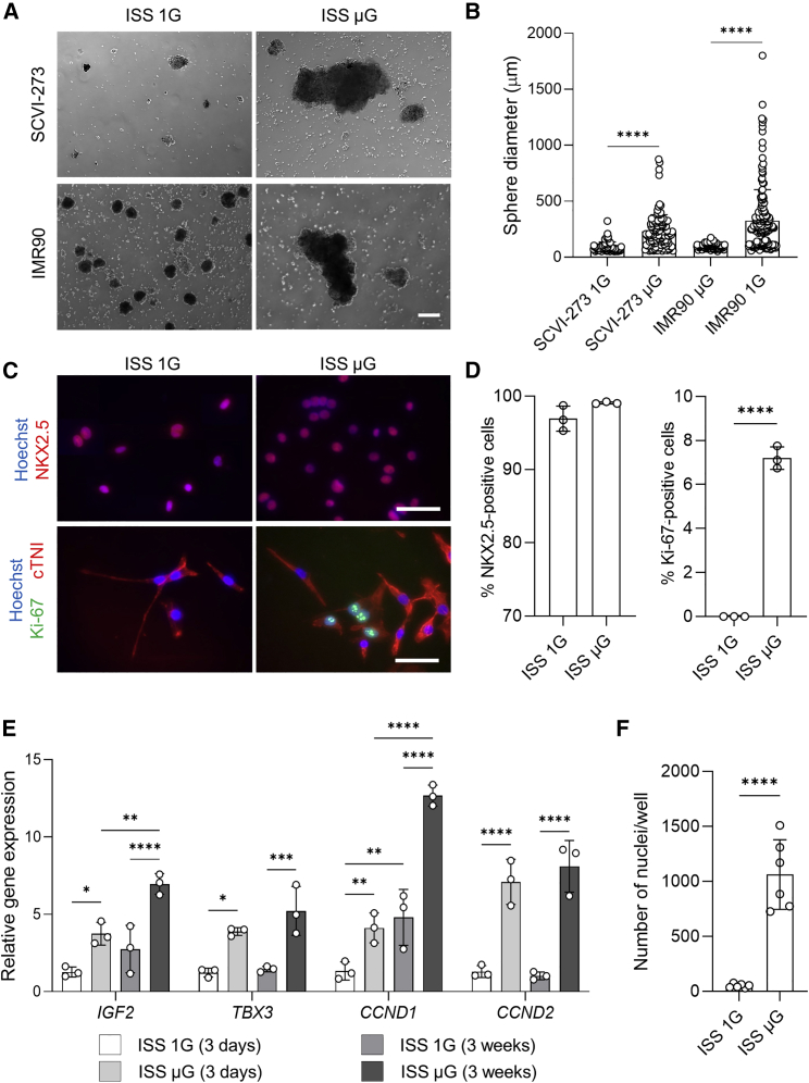
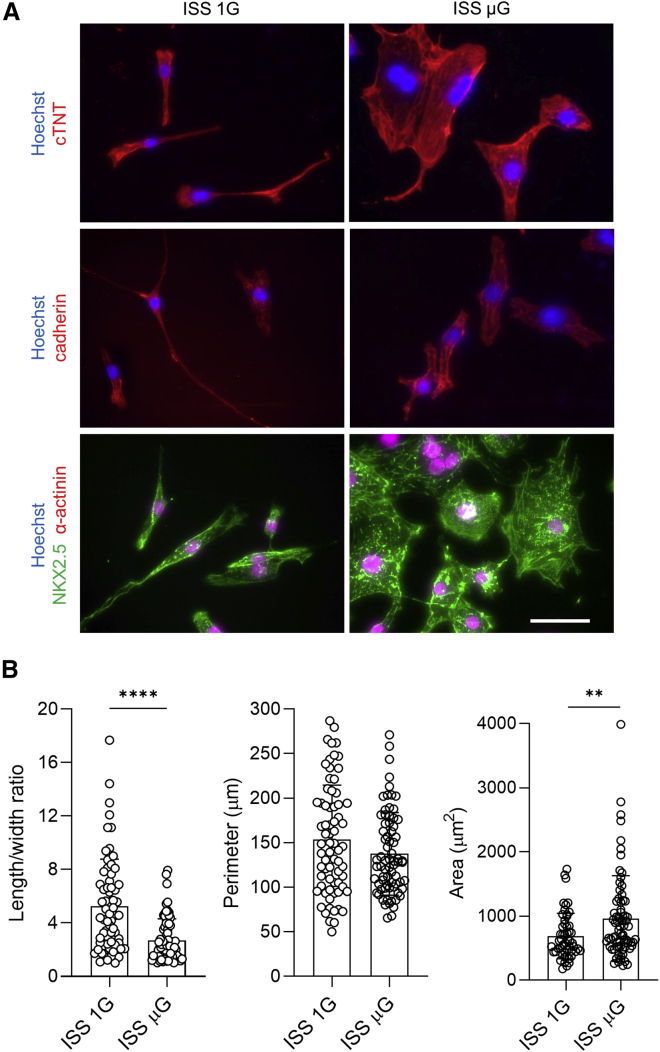
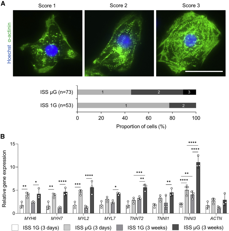
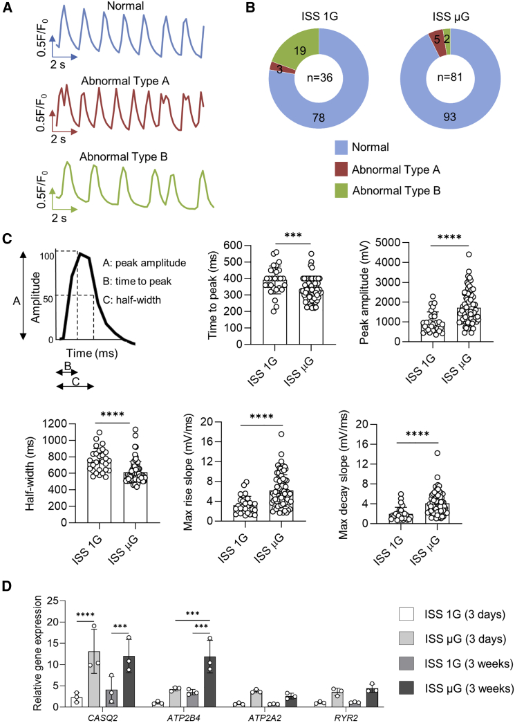
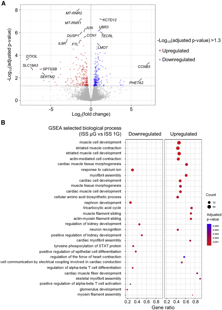
In microgravity, cells undergo profound changes in their properties. However, how human cardiac progenitors respond to space microgravity is unknown. In this study, we evaluated the effect of space microgravity on differentiation of human induced pluripotent stem cell (hiPSC)-derived cardiac progenitors compared with 1G cultures on the International Space Station (ISS). Cryopreserved 3D cardiac progenitors were cultured for 3 weeks on the ISS. Compared with 1G cultures, the microgravity cultures had 3-fold larger sphere sizes, 20-fold higher counts of nuclei, and increased expression of proliferation markers. Highly enriched cardiomyocytes generated in space microgravity showed improved Ca handling and increased expression of contraction-associated genes. Short-term exposure (3 days) of cardiac progenitors to space microgravity upregulated genes involved in cell proliferation, survival, cardiac differentiation, and contraction, consistent with improved microgravity cultures at the late stage. These results indicate that space microgravity increased proliferation of hiPSC-cardiomyocytes, which had appropriate structure and function.
在微重力条件下,细胞的特性会发生深刻变化。然而,人类心脏祖细胞对空间微重力的反应如何尚不清楚。在这项研究中,我们评估了与国际空间站(ISS)上 1G 培养相比,空间微重力对人诱导多能干细胞(hiPSC)衍生的心脏祖细胞分化的影响。冷冻保存的 3D 心脏祖细胞在 ISS 上培养了 3 周。与 1G 培养相比,微重力培养物的球体直径大 3 倍,核数多 20 倍,增殖标志物的表达增加。在空间微重力下生成的高度富集的心肌细胞表现出改善的钙处理和收缩相关基因的表达增加。心脏祖细胞短期(3 天)暴露于空间微重力会上调参与细胞增殖、存活、心脏分化和收缩的基因,这与晚期改善的微重力培养物一致。这些结果表明,空间微重力增加了 hiPSC 心肌细胞的增殖,这些细胞具有适当的结构和功能。