Li Bryan M, Castorina Leonardo V, Valdés Hernández Maria Del C, Clancy Una, Wiseman Stewart J, Sakka Eleni, Storkey Amos J, Jaime Garcia Daniela, Cheng Yajun, Doubal Fergus, Thrippleton Michael T, Stringer Michael, Wardlaw Joanna M
School of Informatics, University of Edinburgh, Edinburgh, United Kingdom.
Centre for Clinical Brain Sciences, University of Edinburgh, Edinburgh, United Kingdom.
Front Comput Neurosci. 2022 Aug 25;16:887633. doi: 10.3389/fncom.2022.887633. eCollection 2022.
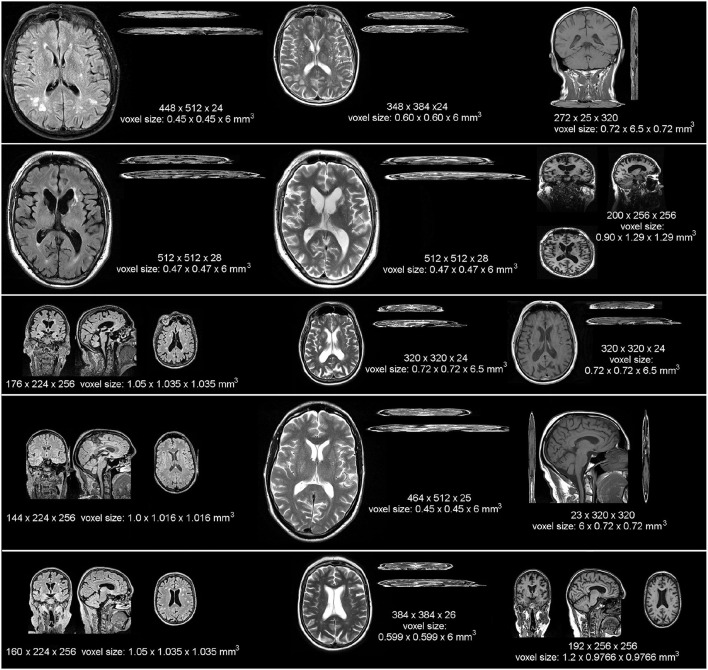
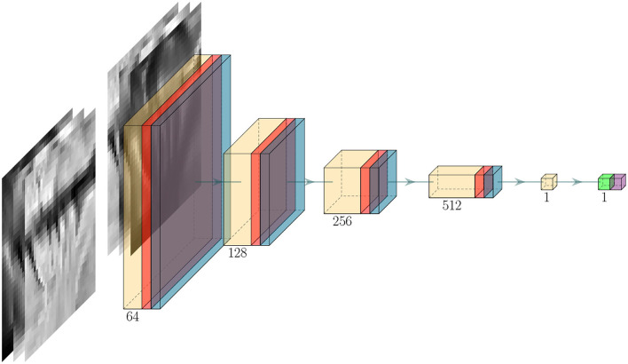
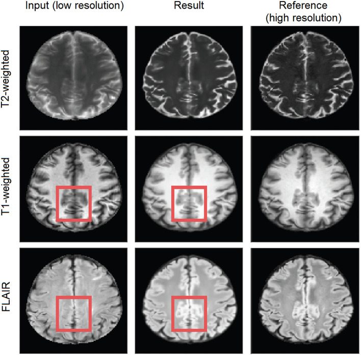
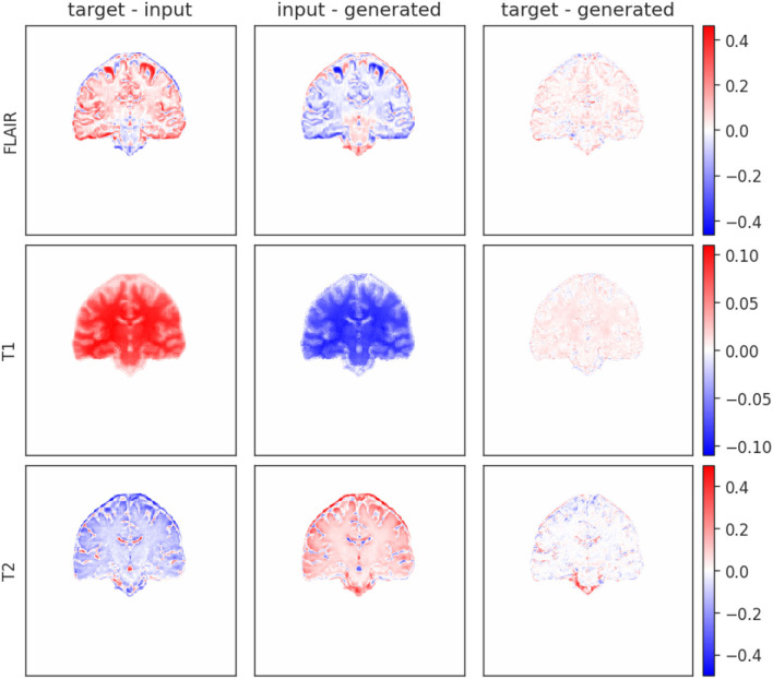
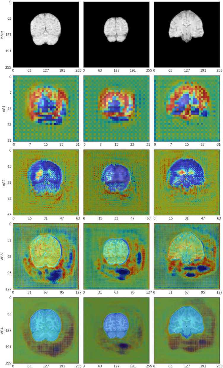
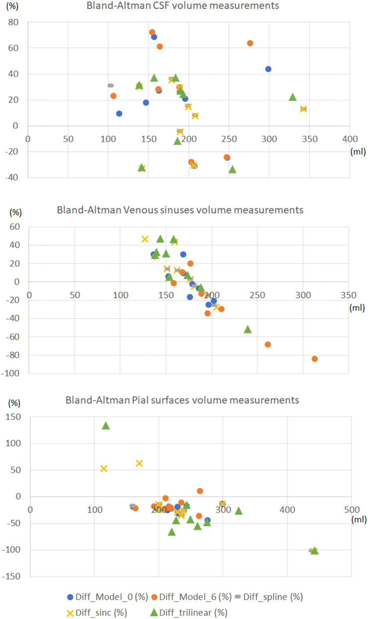
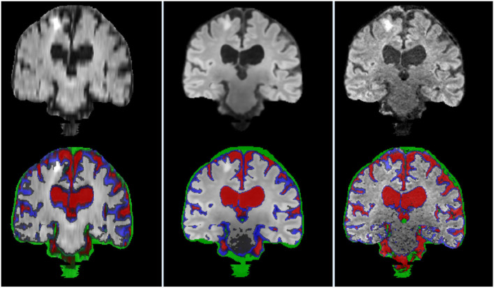
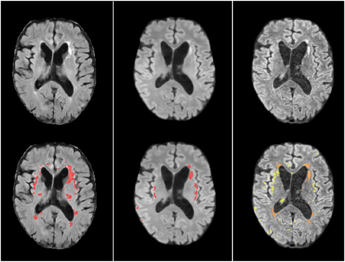
Vast quantities of Magnetic Resonance Images (MRI) are routinely acquired in clinical practice but, to speed up acquisition, these scans are typically of a quality that is sufficient for clinical diagnosis but sub-optimal for large-scale precision medicine, computational diagnostics, and large-scale neuroimaging collaborative research. Here, we present a critic-guided framework to upsample low-resolution (often 2D) MRI full scans to help overcome these limitations. We incorporate feature-importance and self-attention methods into our model to improve the interpretability of this study. We evaluate our framework on paired low- and high-resolution brain MRI structural full scans (i.e., T1-, T2-weighted, and FLAIR sequences are simultaneously input) obtained in clinical and research settings from scanners manufactured by Siemens, Phillips, and GE. We show that the upsampled MRIs are qualitatively faithful to the ground-truth high-quality scans (PSNR = 35.39; MAE = 3.78E-3; NMSE = 4.32E-10; SSIM = 0.9852; mean normal-appearing gray/white matter ratio intensity differences ranging from 0.0363 to 0.0784 for FLAIR, from 0.0010 to 0.0138 for T1-weighted and from 0.0156 to 0.074 for T2-weighted sequences). The automatic raw segmentation of tissues and lesions using the super-resolved images has fewer false positives and higher accuracy than those obtained from interpolated images in protocols represented with more than three sets in the training sample, making our approach a strong candidate for practical application in clinical and collaborative research.
在临床实践中,通常会采集大量的磁共振成像(MRI)数据。然而,为了加快采集速度,这些扫描图像的质量通常仅足以满足临床诊断需求,对于大规模精准医学、计算诊断以及大规模神经影像协作研究而言则不够理想。在此,我们提出了一种由评判器引导的框架,用于对低分辨率(通常为二维)的MRI全扫描图像进行上采样,以帮助克服这些限制。我们将特征重要性和自注意力方法纳入我们的模型,以提高本研究的可解释性。我们在临床和研究环境中,使用西门子、飞利浦和通用电气生产的扫描仪获取的配对低分辨率和高分辨率脑部MRI结构全扫描图像(即同时输入T1加权、T2加权和FLAIR序列)上评估我们的框架。我们表明,上采样后的MRI在质量上与真实的高质量扫描图像相符(PSNR = 35.39;MAE = 3.7e-3;NMSE = 4.32e-10;SSIM = 0.9852;对于FLAIR序列,正常外观的灰质/白质比率强度差异均值范围为0.0363至0.0784,对于T1加权序列为0.0010至0.0138,对于T2加权序列为0.0156至0.074)。使用超分辨率图像进行组织和病变的自动原始分割,与在训练样本中由超过三组表示的协议中从插值图像获得的分割相比,假阳性更少且准确性更高,这使得我们的方法成为临床和协作研究实际应用的有力候选方法。