Jia Yushan, Quan Shuai, Ren Jialiang, Wu Hui, Liu Aishi, Gao Yang, Hao Fene, Yang Zhenxing, Zhang Tong, Hu He
Affiliated Hospital, Inner Mongolia Medical University, Hohhot, China.
Department of Pharmaceuticals Diagnosis, GE Healthcare (China), Shanghai, China.
Front Oncol. 2022 Aug 30;12:974257. doi: 10.3389/fonc.2022.974257. eCollection 2022.
To assess the predictive value of magnetic resonance imaging (MRI) radiomics for progression-free survival (PFS) in patients with prostate cancer (PCa).
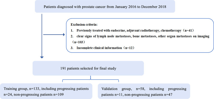
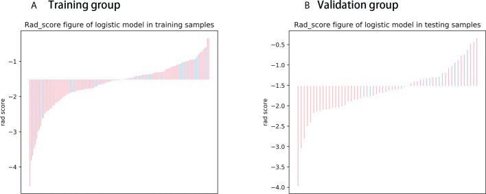
191 patients with prostate cancer confirmed by puncture biopsy or surgical pathology were included in this retrospective study, including 133 in the training group and 58 in the validation group. All patients underwent T2WI and DWI serial scans. Three radiomics models were constructed using univariate logistic regression and Gradient Boosting Decision Tree(GBDT) for feature screening, followed by Cox risk regression to construct a mixed model combining radiomics features and clinicopathological risk factors and to draw a nomogram. The performance of the models was evaluated by receiver operating characteristic curve (ROC), calibration curve and decision curve analysis. The Kaplan-Meier method was applied for survival analysis.
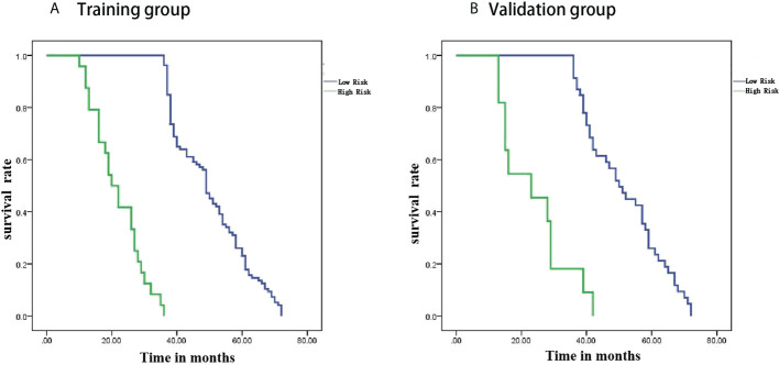
Compared with the radiomics model, the hybrid model consisting of a combination of radiomics features and clinical data performed the best in predicting PFS in PCa patients, with AUCs of 0.926 and 0.917 in the training and validation groups, respectively. Decision curve analysis showed that the radiomics nomogram had good clinical application and the calibration curve proved to have good stability. Survival curves showed that PFS was shorter in the high-risk group than in the low-risk group.
The hybrid model constructed from radiomics and clinical data showed excellent performance in predicting PFS in prostate cancer patients. The nomogram provides a non-invasive diagnostic tool for risk stratification of clinical patients.
评估磁共振成像(MRI)影像组学对前列腺癌(PCa)患者无进展生存期(PFS)的预测价值。
本回顾性研究纳入191例经穿刺活检或手术病理确诊的前列腺癌患者,其中训练组133例,验证组58例。所有患者均接受T2WI和DWI序列扫描。使用单因素逻辑回归和梯度提升决策树(GBDT)进行特征筛选,构建三个影像组学模型,随后采用Cox风险回归构建结合影像组学特征和临床病理危险因素的混合模型并绘制列线图。通过受试者操作特征曲线(ROC)、校准曲线和决策曲线分析评估模型性能。采用Kaplan-Meier法进行生存分析。
与影像组学模型相比,由影像组学特征和临床数据相结合的混合模型在预测PCa患者PFS方面表现最佳,训练组和验证组的AUC分别为0.926和0.917。决策曲线分析表明影像组学列线图具有良好的临床应用价值,校准曲线显示具有良好的稳定性。生存曲线显示高危组的PFS短于低危组。
由影像组学和临床数据构建的混合模型在预测前列腺癌患者PFS方面表现优异。该列线图为临床患者的风险分层提供了一种非侵入性诊断工具。