• 文献检索
  • 文档翻译
  • 深度研究
  • 学术资讯
  • Suppr Zotero 插件Zotero 插件
  • 邀请有礼
  • 套餐&价格
  • 历史记录
应用&插件
Suppr Zotero 插件Zotero 插件浏览器插件Mac 客户端Windows 客户端微信小程序
定价
高级版会员购买积分包购买API积分包
服务
文献检索文档翻译深度研究API 文档MCP 服务
关于我们
关于 Suppr公司介绍联系我们用户协议隐私条款
关注我们

Suppr 超能文献

核心技术专利:CN118964589B侵权必究
粤ICP备2023148730 号-1Suppr @ 2026

文献检索

告别复杂PubMed语法,用中文像聊天一样搜索,搜遍4000万医学文献。AI智能推荐,让科研检索更轻松。

立即免费搜索

文件翻译

保留排版,准确专业,支持PDF/Word/PPT等文件格式,支持 12+语言互译。

免费翻译文档

深度研究

AI帮你快速写综述,25分钟生成高质量综述,智能提取关键信息,辅助科研写作。

立即免费体验

多参数MRI影像组学模型预测卵巢癌术前腹膜癌转移

Multiparameter MRI Radiomics Model Predicts Preoperative Peritoneal Carcinomatosis in Ovarian Cancer.

作者信息

Yu Xiao Yu, Ren Jialiang, Jia Yushan, Wu Hui, Niu Guangming, Liu Aishi, Gao Yang, Hao Fene, Xie Lizhi

机构信息

Affiliated Hospital, Inner Mongolia Medical University, Hohhot, China.

Department of Pharmaceuticals Diagnosis, GE Healthcare (China), Shanghai, China.

出版信息

Front Oncol. 2021 Oct 21;11:765652. doi: 10.3389/fonc.2021.765652. eCollection 2021.

DOI:10.3389/fonc.2021.765652
PMID:34790579
原文链接:https://pmc.ncbi.nlm.nih.gov/articles/PMC8591658/
Abstract

OBJECTIVES

To evaluate the predictive value of radiomics features based on multiparameter magnetic resonance imaging (MP-MRI) for peritoneal carcinomatosis (PC) in patients with ovarian cancer (OC).

METHODS

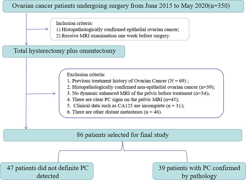
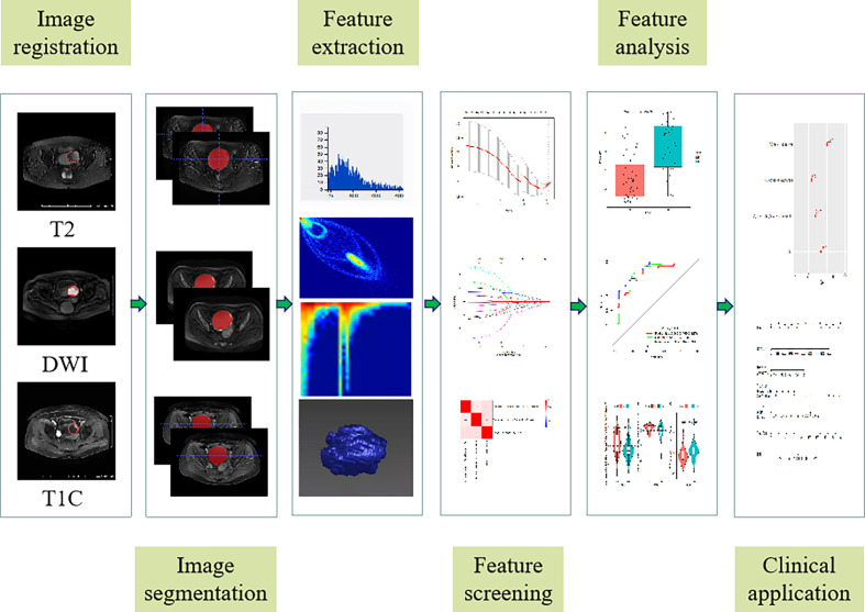
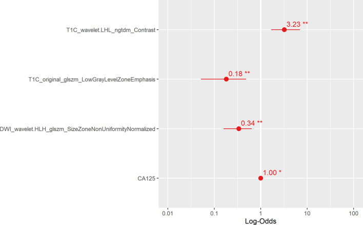
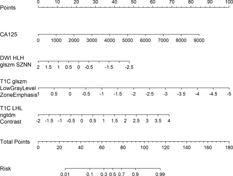
A total of 86 patients with epithelial OC were included in this retrospective study. All patients underwent FS-T2WI, DWI, and DCE-MRI scans, followed by total hysterectomy plus omentectomy. Quantitative imaging features were extracted from preoperative FS-T2WI, DWI, and DCE-MRI images, and feature screening was performed using a minimum redundancy maximum correlation (mRMR) and least absolute shrinkage selection operator (LASSO) methods. Four radiomics models were constructed based on three MRI sequences. Then, combined with radiomics characteristics and clinicopathological risk factors, a multi-factor Logistic regression method was used to construct a radiomics nomogram, and the performance of the radiomics nomogram was evaluated by receiver operating characteristic curve (ROC) curve, calibration curve, and decision curve analysis.

RESULTS

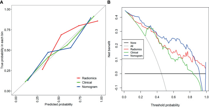
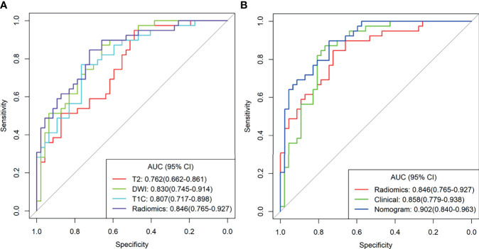
The radiomics model from the MP-MRI combined sequence showed a higher area under the curve (AUC) than the model from FS-T2WI, DWI, and DCE-MRI alone (0.846 . 0.762, 0.830, 0.807, respectively). The radiomics nomogram (AUC=0.902) constructed by combining radiomics characteristics and clinicopathological risk factors showed a better diagnostic effect than the clinical model (AUC=0.858) and the radiomics model (AUC=0.846). The decision curve analysis shows that the radiomics nomogram has good clinical application value, and the calibration curve also proves that it has good stability.

CONCLUSION

Radiomics nomogram based on MP-MRI combined sequence showed good predictive accuracy for PC in patients with OC. This tool can be used to identify peritoneal carcinomatosis in OC patients before surgery.

摘要

目的

评估基于多参数磁共振成像(MP-MRI)的影像组学特征对卵巢癌(OC)患者腹膜癌转移(PC)的预测价值。

方法

本回顾性研究共纳入86例上皮性OC患者。所有患者均接受脂肪抑制T2加权成像(FS-T2WI)、扩散加权成像(DWI)和动态对比增强磁共振成像(DCE-MRI)扫描,随后行全子宫切除术加网膜切除术。从术前FS-T2WI、DWI和DCE-MRI图像中提取定量影像特征,并采用最小冗余最大相关(mRMR)和最小绝对收缩选择算子(LASSO)方法进行特征筛选。基于三个MRI序列构建了四个影像组学模型。然后,结合影像组学特征和临床病理危险因素,采用多因素Logistic回归方法构建影像组学列线图,并通过受试者操作特征曲线(ROC)、校准曲线和决策曲线分析评估影像组学列线图的性能。

结果

MP-MRI联合序列的影像组学模型曲线下面积(AUC)高于单独的FS-T2WI、DWI和DCE-MRI模型(分别为0.846、0.762、0.830、0.807)。结合影像组学特征和临床病理危险因素构建的影像组学列线图(AUC=0.902)比临床模型(AUC=0.858)和影像组学模型(AUC=0.846)具有更好的诊断效果。决策曲线分析表明影像组学列线图具有良好的临床应用价值,校准曲线也证明其具有良好的稳定性。

结论

基于MP-MRI联合序列的影像组学列线图对OC患者的PC具有良好的预测准确性。该工具可用于术前识别OC患者的腹膜癌转移。

https://cdn.ncbi.nlm.nih.gov/pmc/blobs/6c6c/8591658/1af3da7e2ad7/fonc-11-765652-g006.jpg
https://cdn.ncbi.nlm.nih.gov/pmc/blobs/6c6c/8591658/aeabe70aec26/fonc-11-765652-g001.jpg
https://cdn.ncbi.nlm.nih.gov/pmc/blobs/6c6c/8591658/934080deefd7/fonc-11-765652-g002.jpg
https://cdn.ncbi.nlm.nih.gov/pmc/blobs/6c6c/8591658/827791c32b02/fonc-11-765652-g003.jpg
https://cdn.ncbi.nlm.nih.gov/pmc/blobs/6c6c/8591658/a33d747191e3/fonc-11-765652-g004.jpg
https://cdn.ncbi.nlm.nih.gov/pmc/blobs/6c6c/8591658/c6b009fea5ea/fonc-11-765652-g005.jpg
https://cdn.ncbi.nlm.nih.gov/pmc/blobs/6c6c/8591658/1af3da7e2ad7/fonc-11-765652-g006.jpg

相似文献

1
Multiparameter MRI Radiomics Model Predicts Preoperative Peritoneal Carcinomatosis in Ovarian Cancer.多参数MRI影像组学模型预测卵巢癌术前腹膜癌转移
Front Oncol. 2021 Oct 21;11:765652. doi: 10.3389/fonc.2021.765652. eCollection 2021.
2
[Preoperative prediction of HER-2 expression status in breast cancer based on MRI radiomics model].基于MRI影像组学模型的乳腺癌HER-2表达状态术前预测
Zhonghua Zhong Liu Za Zhi. 2024 May 23;46(5):428-437. doi: 10.3760/cma.j.cn112152-20230816-00086.
3
Prediction of Lymphovascular Space Invision in Endometrial Cancer based on Multi-parameter MRI Radiomics Model.基于多参数MRI影像组学模型预测子宫内膜癌的脉管间隙浸润
Curr Med Imaging. 2024 Mar 19. doi: 10.2174/0115734056266366231219111246.
4
An MRI-Based Radiomics Model for Predicting the Benignity and Malignancy of BI-RADS 4 Breast Lesions.一种基于磁共振成像的影像组学模型用于预测乳腺影像报告和数据系统(BI-RADS)4类乳腺病变的良恶性
Front Oncol. 2022 Jan 28;11:733260. doi: 10.3389/fonc.2021.733260. eCollection 2021.
5
Multiparametric MRI-based radiomics nomogram for differentiation of primary mucinous ovarian cancer from metastatic ovarian cancer.基于多参数磁共振成像的影像组学列线图用于鉴别原发性黏液性卵巢癌与转移性卵巢癌
Abdom Radiol (NY). 2025 Feb;50(2):1018-1028. doi: 10.1007/s00261-024-04542-y. Epub 2024 Aug 31.
6
[Prediction of platinum-based chemotherapy sensitivity for epithelial ovarian cancer by multi-sequence MRI-based radiomic nomogram].基于多序列MRI的影像组学列线图预测上皮性卵巢癌铂类化疗敏感性
Zhonghua Yi Xue Za Zhi. 2022 Jan 18;102(3):201-208. doi: 10.3760/cma.j.cn112137-20210816-01844.
7
Radiomic Evaluations of the Diagnostic Performance of DM, DBT, DCE MRI, DWI, and Their Combination for the Diagnosisof Breast Cancer.数字乳腺摄影(DM)、数字乳腺断层摄影(DBT)、动态对比增强磁共振成像(DCE MRI)、扩散加权成像(DWI)及其联合应用对乳腺癌诊断性能的影像组学评估
Front Oncol. 2021 Sep 10;11:725922. doi: 10.3389/fonc.2021.725922. eCollection 2021.
8
MRI-based radiomics analysis to predict preoperative lymph node metastasis in papillary thyroid carcinoma.基于磁共振成像的影像组学分析预测甲状腺乳头状癌术前淋巴结转移
Gland Surg. 2020 Oct;9(5):1214-1226. doi: 10.21037/gs-20-479.
9
Biparametric magnetic resonance imaging-based radiomics features for prediction of lymphovascular invasion in rectal cancer.基于双参数磁共振成像的放射组学特征预测直肠癌的血管淋巴管侵犯。
BMC Cancer. 2023 Jan 18;23(1):61. doi: 10.1186/s12885-023-10534-w.
10
Preoperative prediction of parametrial invasion in early-stage cervical cancer with MRI-based radiomics nomogram.基于 MRI 影像组学列线图预测早期宫颈癌的宫旁侵犯
Eur Radiol. 2020 Jun;30(6):3585-3593. doi: 10.1007/s00330-019-06655-1. Epub 2020 Feb 17.

引用本文的文献

1
Development and validation of multi-sequence magnetic resonance imaging radiomics models for predicting tumor response to radiotherapy in locally advanced non-small cell lung cancer.用于预测局部晚期非小细胞肺癌放疗肿瘤反应的多序列磁共振成像放射组学模型的开发与验证
J Thorac Dis. 2025 Mar 31;17(3):1684-1697. doi: 10.21037/jtd-2025-142. Epub 2025 Mar 14.
2
Radiomics and radiogenomics: extracting more information from medical images for the diagnosis and prognostic prediction of ovarian cancer.放射组学和放射基因组学:从医学影像中提取更多信息用于卵巢癌的诊断和预后预测。
Mil Med Res. 2024 Dec 14;11(1):77. doi: 10.1186/s40779-024-00580-1.
3

本文引用的文献

1
Predicting postoperative peritoneal metastasis in gastric cancer with serosal invasion using a collagen nomogram.基于胶原列线图预测伴有浆膜侵犯的胃癌患者术后腹膜转移
Nat Commun. 2021 Jan 8;12(1):179. doi: 10.1038/s41467-020-20429-0.
2
MR imaging of epithelial ovarian cancer: a combined model to predict histologic subtypes.磁共振成像在卵巢上皮性癌中的应用:一种预测组织学亚型的联合模型。
Eur Radiol. 2020 Nov;30(11):5815-5825. doi: 10.1007/s00330-020-06993-5. Epub 2020 Jun 13.
3
Bone Marrow and Tumor Radiomics at F-FDG PET/CT: Impact on Outcome Prediction in Non-Small Cell Lung Cancer.
Intratumoral and peritumoral MRI-based radiomics for predicting extrapelvic peritoneal metastasis in epithelial ovarian cancer.
基于磁共振成像(MRI)的肿瘤内和瘤周影像组学预测上皮性卵巢癌盆腔外腹膜转移
Insights Imaging. 2024 Nov 22;15(1):281. doi: 10.1186/s13244-024-01855-w.
4
Advances in ovarian cancer radiomics: a bibliometric analysis from 2010 to 2024.卵巢癌影像组学的进展:2010年至2024年的文献计量分析
Front Oncol. 2024 Oct 1;14:1456932. doi: 10.3389/fonc.2024.1456932. eCollection 2024.
5
Predicting CD27 expression and clinical prognosis in serous ovarian cancer using CT-based radiomics.基于 CT 影像组学预测浆液性卵巢癌中 CD27 的表达和临床预后
J Ovarian Res. 2024 Jun 22;17(1):131. doi: 10.1186/s13048-024-01456-7.
6
Advances in artificial intelligence for the diagnosis and treatment of ovarian cancer (Review).人工智能在卵巢癌诊断和治疗中的进展(综述)。
Oncol Rep. 2024 Mar;51(3). doi: 10.3892/or.2024.8705. Epub 2024 Jan 19.
7
Imaging Evaluation of Peritoneal Metastasis: Current and Promising Techniques.腹膜转移瘤的影像学评估:现有及有潜力的技术。
Korean J Radiol. 2024 Jan;25(1):86-102. doi: 10.3348/kjr.2023.0840.
8
Imaging of Peritoneal Carcinomatosis in Advanced Ovarian Cancer: CT, MRI, Radiomic Features and Resectability Criteria.晚期卵巢癌腹膜转移瘤的影像学检查:CT、MRI、影像组学特征及可切除性标准
Cancers (Basel). 2023 Dec 13;15(24):5827. doi: 10.3390/cancers15245827.
9
Ovarian cancer beyond imaging: integration of AI and multiomics biomarkers.卵巢癌的影像学之外:人工智能与多组学生物标志物的整合。
Eur Radiol Exp. 2023 Sep 13;7(1):50. doi: 10.1186/s41747-023-00364-7.
10
A systematic review and meta-analysis of CT and MRI radiomics in ovarian cancer: methodological issues and clinical utility.卵巢癌CT和MRI影像组学的系统评价与Meta分析:方法学问题与临床应用
Insights Imaging. 2023 Jul 3;14(1):117. doi: 10.1186/s13244-023-01464-z.
骨髓和肿瘤 CT 体层摄影术 F-FDG 摄取的放射组学:对非小细胞肺癌预后预测的影响。
Radiology. 2019 Nov;293(2):451-459. doi: 10.1148/radiol.2019190357. Epub 2019 Sep 17.
4
Radiomics analysis using contrast-enhanced CT for preoperative prediction of occult peritoneal metastasis in advanced gastric cancer.基于增强 CT 的影像组学分析对进展期胃癌隐匿性腹膜转移的术前预测
Eur Radiol. 2020 Jan;30(1):239-246. doi: 10.1007/s00330-019-06368-5. Epub 2019 Aug 5.
5
Radiomics Analysis of Gadoxetic Acid-enhanced MRI for Staging Liver Fibrosis.钆塞酸二钠增强MRI对肝纤维化分期的影像组学分析
Radiology. 2019 Jul;292(1):269. doi: 10.1148/radiol.2019194012.
6
Development and validation of an individualized nomogram to identify occult peritoneal metastasis in patients with advanced gastric cancer.开发和验证一种个体化列线图以识别晚期胃癌患者隐匿性腹膜转移。
Ann Oncol. 2019 Mar 1;30(3):431-438. doi: 10.1093/annonc/mdz001.
7
Combined Benefit of Quantitative Three-Compartment Breast Image Analysis and Mammography Radiomics in the Classification of Breast Masses in a Clinical Data Set.定量三腔乳腺图像分析与乳腺 X 线摄影组学在临床数据集乳腺肿块分类中的联合获益。
Radiology. 2019 Mar;290(3):621-628. doi: 10.1148/radiol.2018180608. Epub 2018 Dec 11.
8
A radiomics approach to assess tumour-infiltrating CD8 cells and response to anti-PD-1 or anti-PD-L1 immunotherapy: an imaging biomarker, retrospective multicohort study.一种基于放射组学的方法来评估肿瘤浸润 CD8 细胞与抗 PD-1 或抗 PD-L1 免疫治疗反应的关系:一项影像学生物标志物、回顾性多队列研究。
Lancet Oncol. 2018 Sep;19(9):1180-1191. doi: 10.1016/S1470-2045(18)30413-3. Epub 2018 Aug 14.
9
Comparison between multi-slice spiral CT and magnetic resonance imaging in the diagnosis of peritoneal metastasis in primary ovarian carcinoma.多层螺旋CT与磁共振成像在原发性卵巢癌腹膜转移诊断中的比较
Onco Targets Ther. 2018 Feb 28;11:1087-1094. doi: 10.2147/OTT.S147700. eCollection 2018.
10
Cancer statistics, 2018.癌症统计数据,2018 年。
CA Cancer J Clin. 2018 Jan;68(1):7-30. doi: 10.3322/caac.21442. Epub 2018 Jan 4.