Zhou Yi, Liu Xiaoyan, She Hongjiang, Wang Rui, Bai Fan, Xiang Bingyan
Department of Orthopaedics, Third Affiliated Hospital of Zunyi Medical University (The First People's Hospital of Zunyi City), Zunyi 563000, China.
Regen Ther. 2022 Sep 2;21:307-321. doi: 10.1016/j.reth.2022.08.006. eCollection 2022 Dec.
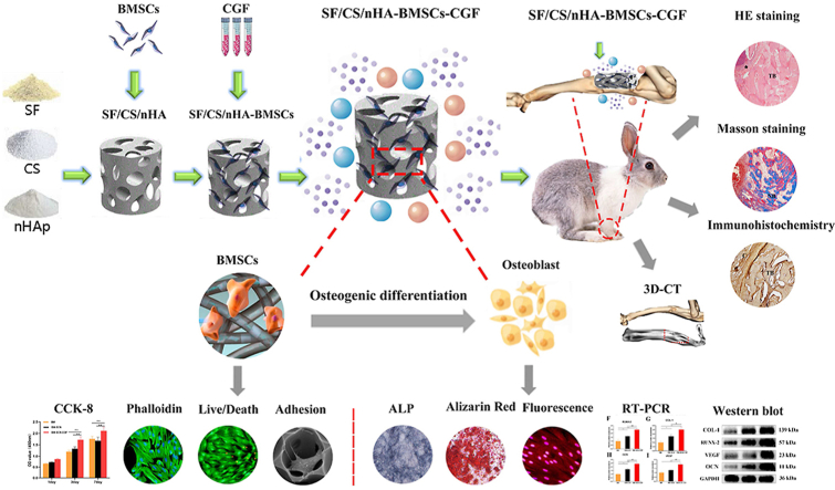
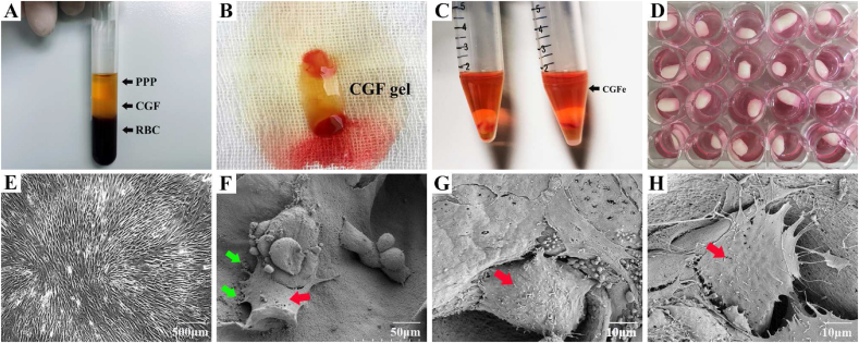
With the goal of increasing the translational efficiency of bone tissue engineering for practical clinical applications, biomimetic composite scaffolds combined with autologous endogenous growth factors for repairing bone defects have become a current research hotspot. In this study, we prepared a silk fibroin/chitosan/nanohydroxyapatite (SF/CS/nHA) composite biomimetic scaffold and then combined it with autologous concentrated growth factor (CGF) to explore the effect of this combination on the proliferation and osteogenic differentiation of bone marrow mesenchymal stem cells (BMSCs) and the efficiency of repairing critical radial defects.
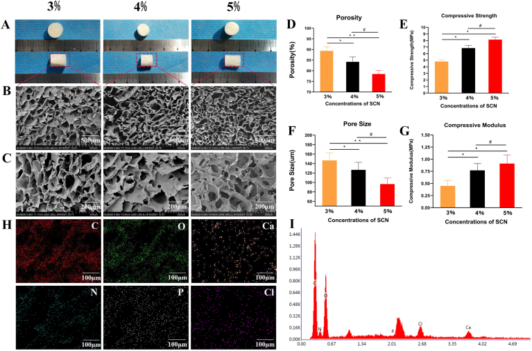
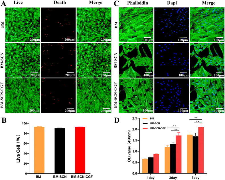
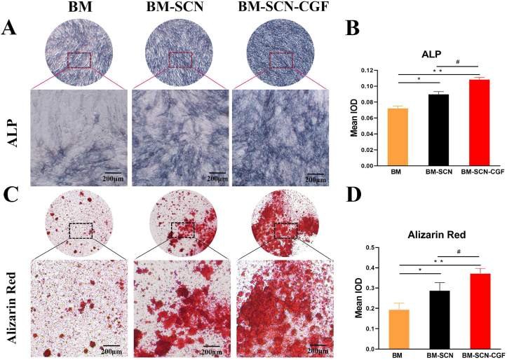
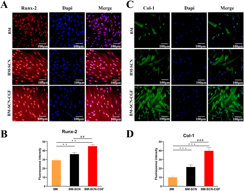
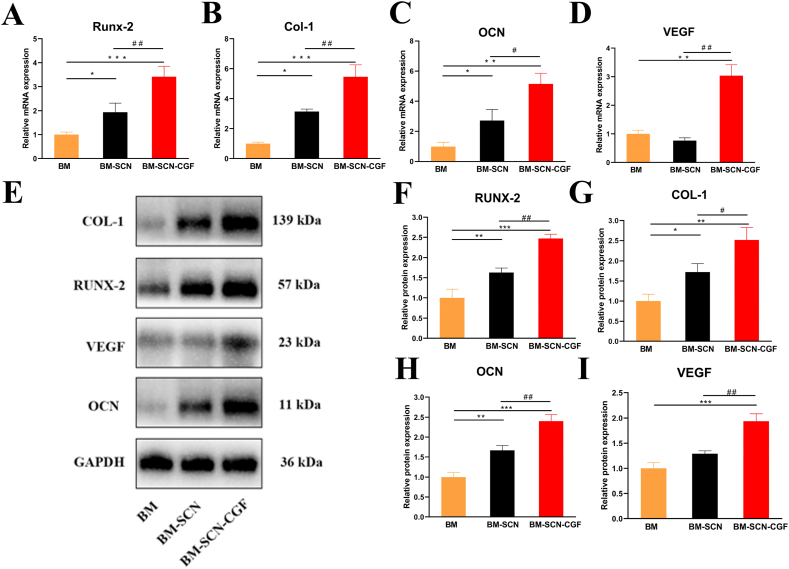
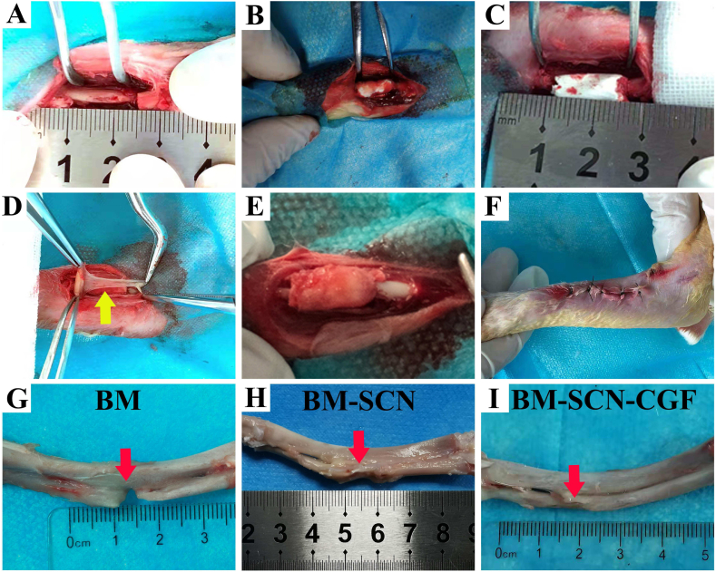
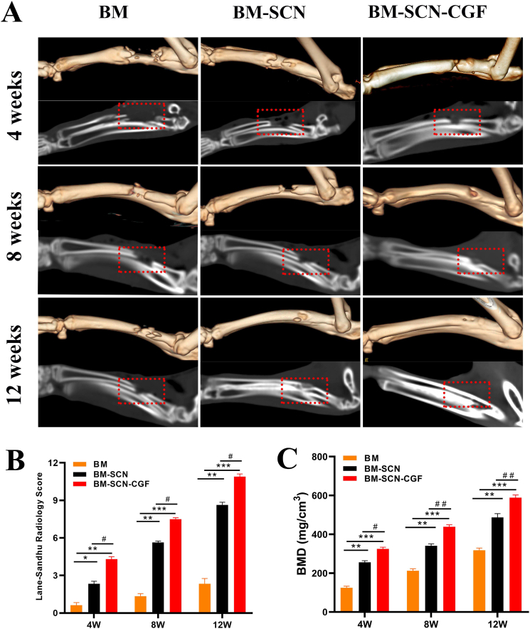
Three kinds of SF/CS/nHA composite biomimetic scaffolds with mass fractions of 3%, 4%, and 5% were prepared by vacuum freeze-drying and chemical cross-linking methods, and the characteristics of the scaffolds were evaluated. In vitro, BMSCs were seeded on SF/CS/nHA scaffolds, and then CGF was added. The morphology and proliferation of BMSCs were evaluated by live-dead staining, phalloidin staining, and CCK-8 assays. ALP staining, alizarin red staining, cellular immunofluorescence, RT-PCR, and Western blotting were used to detect the osteogenic differentiation of BMSCs. In vivo, a rabbit radius critical bone defect model was constructed, and the SF/CS/nHA-BMSC scaffold cell complex combined with CGF was implanted. The effect on bone defect repair was evaluated by 3D CT scanning, HE staining, Masson staining, and immunohistochemistry.
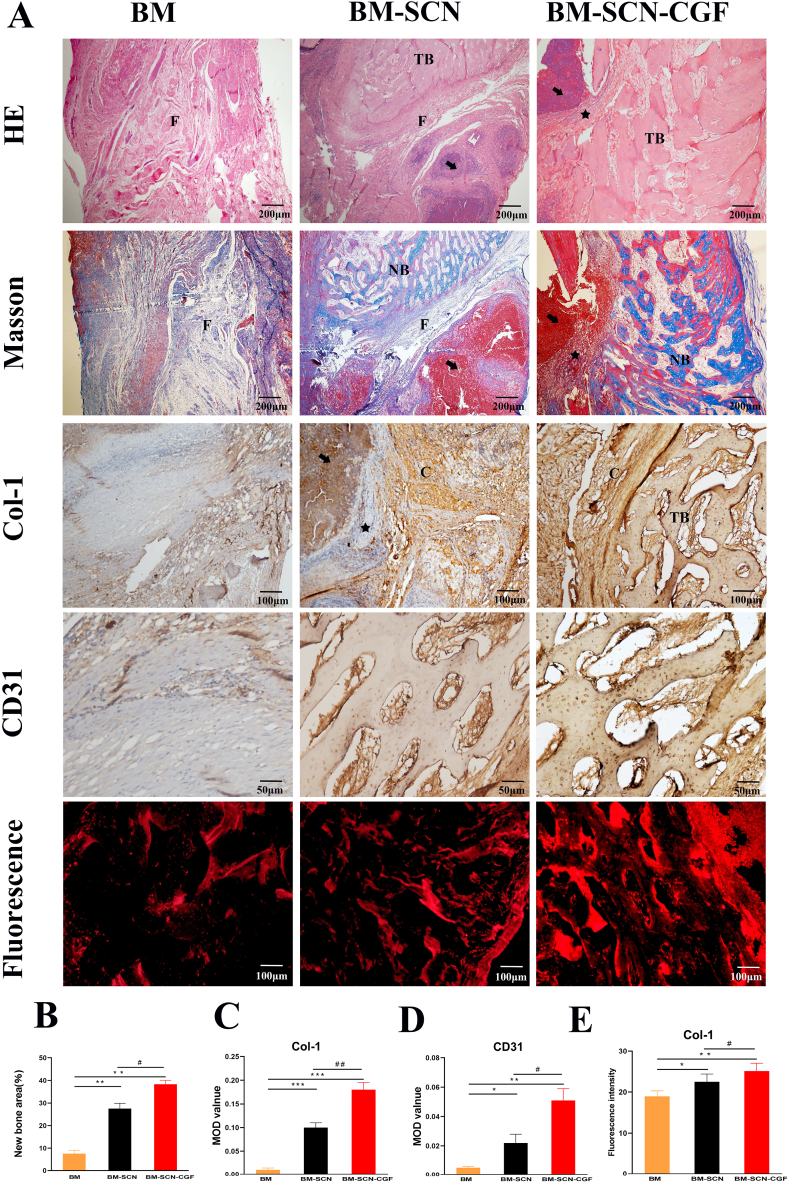
The characteristics of 4% SF/CS/nHA were the most suitable for repairing bone defects. In vitro, the SF/CS/nHA combined CGF group showed better adhesion, cell morphology, proliferation, and osteogenic differentiation of BMSCs than the other groups (P < 0.05 for all). In vivo imaging examination and histological analysis demonstrated that the SF/CS/nHA scaffold combined with CGF had better efficiency in bone defect repair than the other scaffolds (P < 0.05 for all).
A SF/CS/nHA composite biomimetic bone scaffold combined with autologous CGF promoted the proliferation and osteogenic differentiation of BMSCs in vitro and improved the repair efficiency of critical bone defects in vivo. This combination may have the potential for clinical translation due to its excellent biocompatibility.
为提高骨组织工程在实际临床应用中的转化效率,将生物仿生复合支架与自体源性生长因子相结合用于修复骨缺损已成为当前的研究热点。在本研究中,我们制备了丝素蛋白/壳聚糖/纳米羟基磷灰石(SF/CS/nHA)复合生物仿生支架,然后将其与自体浓缩生长因子(CGF)相结合,以探讨这种组合对骨髓间充质干细胞(BMSCs)增殖和成骨分化的影响以及修复桡骨临界性缺损的效率。
采用真空冷冻干燥和化学交联法制备质量分数分别为3%、4%和5%的三种SF/CS/nHA复合生物仿生支架,并对支架特性进行评估。在体外,将BMSCs接种于SF/CS/nHA支架上,然后添加CGF。通过活死染色、鬼笔环肽染色和CCK-8检测评估BMSCs的形态和增殖情况。采用碱性磷酸酶(ALP)染色、茜素红染色、细胞免疫荧光、逆转录-聚合酶链反应(RT-PCR)和蛋白质免疫印迹法检测BMSCs的成骨分化情况。在体内,构建兔桡骨临界性骨缺损模型,植入SF/CS/nHA-BMSC支架细胞复合物并联合CGF。通过三维计算机断层扫描(3D CT)、苏木精-伊红(HE)染色、马松(Masson)染色和免疫组织化学评估对骨缺损修复的效果。
4% SF/CS/nHA的特性最适合修复骨缺损。在体外,与其他组相比,SF/CS/nHA联合CGF组的BMSCs表现出更好的黏附、细胞形态、增殖和成骨分化(所有P<0.05)。体内成像检查和组织学分析表明,与其他支架相比,SF/CS/nHA支架联合CGF在骨缺损修复方面具有更高的效率(所有P<0.05)。
SF/CS/nHA复合生物仿生骨支架联合自体CGF在体外促进了BMSCs的增殖和成骨分化,并在体内提高了临界性骨缺损的修复效率。由于其优异的生物相容性,这种组合可能具有临床转化的潜力。