• 文献检索
  • 文档翻译
  • 深度研究
  • 学术资讯
  • Suppr Zotero 插件Zotero 插件
  • 邀请有礼
  • 套餐&价格
  • 历史记录
应用&插件
Suppr Zotero 插件Zotero 插件浏览器插件Mac 客户端Windows 客户端微信小程序
定价
高级版会员购买积分包购买API积分包
服务
文献检索文档翻译深度研究API 文档MCP 服务
关于我们
关于 Suppr公司介绍联系我们用户协议隐私条款
关注我们

Suppr 超能文献

核心技术专利:CN118964589B侵权必究
粤ICP备2023148730 号-1Suppr @ 2026

文献检索

告别复杂PubMed语法,用中文像聊天一样搜索,搜遍4000万医学文献。AI智能推荐,让科研检索更轻松。

立即免费搜索

文件翻译

保留排版,准确专业,支持PDF/Word/PPT等文件格式,支持 12+语言互译。

免费翻译文档

深度研究

AI帮你快速写综述,25分钟生成高质量综述,智能提取关键信息,辅助科研写作。

立即免费体验

基于扫频源光学相干断层扫描图像的像素级深度学习模型检测白内障的建立与验证。

Development and validation of a pixel wise deep learning model to detect cataract on swept-source optical coherence tomography images.

机构信息

Department of Ophthalmology, Rothschild Foundation, 25 Rue Manin, Paris 75019, France.

Department of Ophthalmology, Rothschild Foundation, 25 Rue Manin, Paris 75019, France.

出版信息

J Optom. 2022;15 Suppl 1(Suppl 1):S43-S49. doi: 10.1016/j.optom.2022.08.003. Epub 2022 Oct 10.

DOI:10.1016/j.optom.2022.08.003
PMID:36229338
原文链接:https://pmc.ncbi.nlm.nih.gov/articles/PMC9732477/
Abstract

PURPOSE

The diagnosis of cataract is mostly clinical and there is a lack of objective and specific tool to detect and grade it automatically. The goal of this study was to develop and validate a deep learning model to detect and localize cataract on Swept Source Optical Coherance Tomography (SS-OCT) images.

METHODS

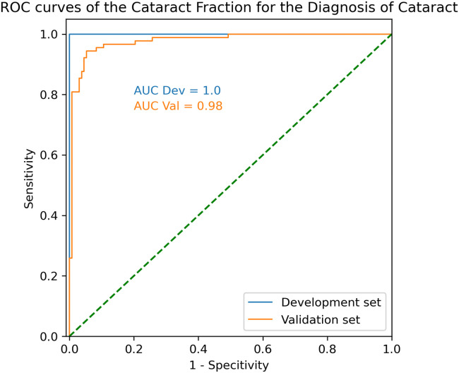
We trained a convolutional network to detect cataract at the pixel level from 504 SS-OCT images of clear lens and cataract patients. The model was then validated on 1326 different images of 114 patients. The output of the model is a map repreenting the probability of cataract for each pixel of the image. We calculated the Cataract Fraction (CF), defined as the number of pixel classified as "cataract" divided by the number of pixel representing the lens for each image. Receiver Operating Characteristic Curves were plotted. Area Under the Curve (ROC AUC) sensitivity and specitivity to detect cataract were calculated.

RESULTS

In the validsation set, mean CF was 0.024 ± 0.077 and 0.479 ± 0.230 (p < 0.001). ROC AUC was 0.98 with an optimal CF threshold of 0.14. Using that threshold, sensitivity and specificity to detect cataract were 94.4% and 94.7%, respectively.

CONCLUSION

We developed an automatic detection tool for cataract on SS-OCT images. Probability maps of cataract on the images provide an additional tool to help the physician in its diagnosis and surgical planning.

摘要

目的

白内障的诊断主要是临床诊断,缺乏客观和特定的工具来自动检测和分级。本研究的目的是开发和验证一种深度学习模型,以自动检测和定位扫频源光学相干断层扫描(SS-OCT)图像中的白内障。

方法

我们使用卷积网络从 504 张清晰晶状体和白内障患者的 SS-OCT 图像中逐像素地训练模型以检测白内障。然后,我们在 114 名患者的 1326 张不同图像上验证了该模型。模型的输出是一张代表图像中每个像素白内障概率的地图。我们计算了白内障分数(CF),定义为被归类为“白内障”的像素数除以代表图像中晶状体的像素数。绘制了受试者工作特征曲线。计算了用于检测白内障的曲线下面积(ROC AUC)的敏感性和特异性。

结果

在验证集中,平均 CF 为 0.024±0.077 和 0.479±0.230(p<0.001)。ROC AUC 为 0.98,最佳 CF 阈值为 0.14。使用该阈值,检测白内障的敏感性和特异性分别为 94.4%和 94.7%。

结论

我们开发了一种用于 SS-OCT 图像中白内障的自动检测工具。图像上的白内障概率图提供了一种额外的工具,有助于医生进行诊断和手术规划。

https://cdn.ncbi.nlm.nih.gov/pmc/blobs/ca6f/9732477/a720170f50f9/gr3.jpg
https://cdn.ncbi.nlm.nih.gov/pmc/blobs/ca6f/9732477/e4c049d87e31/gr1.jpg
https://cdn.ncbi.nlm.nih.gov/pmc/blobs/ca6f/9732477/6adf95476ce7/gr2.jpg
https://cdn.ncbi.nlm.nih.gov/pmc/blobs/ca6f/9732477/a720170f50f9/gr3.jpg
https://cdn.ncbi.nlm.nih.gov/pmc/blobs/ca6f/9732477/e4c049d87e31/gr1.jpg
https://cdn.ncbi.nlm.nih.gov/pmc/blobs/ca6f/9732477/6adf95476ce7/gr2.jpg
https://cdn.ncbi.nlm.nih.gov/pmc/blobs/ca6f/9732477/a720170f50f9/gr3.jpg

相似文献

1
Development and validation of a pixel wise deep learning model to detect cataract on swept-source optical coherence tomography images.基于扫频源光学相干断层扫描图像的像素级深度学习模型检测白内障的建立与验证。
J Optom. 2022;15 Suppl 1(Suppl 1):S43-S49. doi: 10.1016/j.optom.2022.08.003. Epub 2022 Oct 10.
2
Average lens density quantification with swept-source optical coherence tomography: optimized, automated cataract grading technique.应用扫频源光相干断层扫描进行平均晶状体密度定量分析:优化的、自动化的白内障分级技术。
J Cataract Refract Surg. 2019 Dec;45(12):1746-1752. doi: 10.1016/j.jcrs.2019.07.033.
3
Corneal Edema Visualization With Optical Coherence Tomography Using Deep Learning: Proof of Concept.基于深度学习的光学相干断层扫描术对角膜水肿的可视化:概念验证。
Cornea. 2021 Oct 1;40(10):1267-1275. doi: 10.1097/ICO.0000000000002640.
4
Three-Dimensional Cataract Crystalline Lens Imaging With Swept-Source Optical Coherence Tomography.扫频源光学相干断层扫描的三维白内障晶状体成像。
Invest Ophthalmol Vis Sci. 2018 Feb 1;59(2):897-903. doi: 10.1167/iovs.17-23596.
5
Accuracy of swept source biometry device in detecting macular diseases compared with swept source OCT.扫频源生物测量仪与扫频源 OCT 检测黄斑疾病的准确性比较。
BMC Res Notes. 2023 Dec 19;16(1):376. doi: 10.1186/s13104-023-06641-3.
6
Cataract quantification using swept-source optical coherence tomography.应用扫频源光学相干断层扫描对白内障进行定量分析。
J Cataract Refract Surg. 2018 Dec;44(12):1478-1481. doi: 10.1016/j.jcrs.2018.08.009. Epub 2018 Nov 2.
7
OCT-based deep learning algorithm for the evaluation of treatment indication with anti-vascular endothelial growth factor medications.基于光学相干断层扫描的深度学习算法用于评估抗血管内皮生长因子药物的治疗指征
Graefes Arch Clin Exp Ophthalmol. 2018 Jan;256(1):91-98. doi: 10.1007/s00417-017-3839-y. Epub 2017 Nov 10.
8
Accuracy of Swept-Source Optical Coherence Tomography and Ultrasound Biomicroscopy for Evaluation of Posterior Lens Capsule in Traumatic Cataract.应用扫频源光学相干断层扫描和超声生物显微镜评估外伤性白内障后囊膜的准确性。
Am J Ophthalmol. 2020 Aug;216:55-58. doi: 10.1016/j.ajo.2020.03.030. Epub 2020 Apr 2.
9
Efficacy of Swept-source Optical Coherence Tomography in Axial Length Measurement for Advanced Cataract Patients.扫频源光学相干断层扫描在高度近视白内障患者眼轴测量中的疗效。
Optom Vis Sci. 2020 Mar;97(3):186-191. doi: 10.1097/OPX.0000000000001491.
10
Fully automated detection of retinal disorders by image-based deep learning.基于图像的深度学习技术对视网膜疾病进行全自动检测。
Graefes Arch Clin Exp Ophthalmol. 2019 Mar;257(3):495-505. doi: 10.1007/s00417-018-04224-8. Epub 2019 Jan 4.

引用本文的文献

1
The Clinical Usefulness of Evaluating the Lens and Intraocular Lenses Using Optical Coherence Tomography: An Updated Literature Review.使用光学相干断层扫描评估晶状体和人工晶状体的临床实用性:最新文献综述
J Clin Med. 2024 Nov 22;13(23):7070. doi: 10.3390/jcm13237070.
2
Artificial Intelligence for Anterior Segment Diseases: A Review of Potential Developments and Clinical Applications.用于眼前节疾病的人工智能:潜在发展与临床应用综述
Ophthalmol Ther. 2023 Jun;12(3):1439-1455. doi: 10.1007/s40123-023-00690-4. Epub 2023 Mar 8.
3
Journal of Optometry Impact Factor: it's been a long way to here.

本文引用的文献

1
Mixed pyramid attention network for nuclear cataract classification based on anterior segment OCT images.基于眼前节光学相干断层扫描(OCT)图像的混合金字塔注意力网络用于核性白内障分类
Health Inf Sci Syst. 2022 Mar 25;10(1):3. doi: 10.1007/s13755-022-00170-2. eCollection 2022 Dec.
2
DeepLensNet: Deep Learning Automated Diagnosis and Quantitative Classification of Cataract Type and Severity.DeepLensNet:深度学习自动诊断和白内障类型及严重程度的定量分类。
Ophthalmology. 2022 May;129(5):571-584. doi: 10.1016/j.ophtha.2021.12.017. Epub 2022 Jan 3.
3
GLA-Net: A global-local attention network for automatic cataract classification.
《验光杂志》影响因子:走到今天历经漫长历程。
J Optom. 2023 Jan-Mar;16(1):1-2. doi: 10.1016/j.optom.2022.12.001.
GLA-Net:用于自动白内障分类的全局-局部注意力网络。
J Biomed Inform. 2021 Dec;124:103939. doi: 10.1016/j.jbi.2021.103939. Epub 2021 Nov 6.
4
Lens Opacities Classification System III-based artificial intelligence program for automatic cataract grading.基于 Lens Opacities Classification System III 的人工智能程序用于自动白内障分级。
J Cataract Refract Surg. 2022 May 1;48(5):528-534. doi: 10.1097/j.jcrs.0000000000000790.
5
Corneal Edema Visualization With Optical Coherence Tomography Using Deep Learning: Proof of Concept.基于深度学习的光学相干断层扫描术对角膜水肿的可视化:概念验证。
Cornea. 2021 Oct 1;40(10):1267-1275. doi: 10.1097/ICO.0000000000002640.
6
Assessment of Visual Quality in Eyes with Forme Fruste Keratoconus and Mild and Moderate Keratoconus Based on Optical Quality Analysis System II Parameters.基于光学质量分析系统II参数评估亚临床圆锥角膜及轻、中度圆锥角膜患者的视觉质量
J Ophthalmol. 2020 Feb 29;2020:7505016. doi: 10.1155/2020/7505016. eCollection 2020.
7
Average lens density quantification with swept-source optical coherence tomography: optimized, automated cataract grading technique.应用扫频源光相干断层扫描进行平均晶状体密度定量分析:优化的、自动化的白内障分级技术。
J Cataract Refract Surg. 2019 Dec;45(12):1746-1752. doi: 10.1016/j.jcrs.2019.07.033.
8
Volumetric macro- and micro-scale assessment of crystalline lens opacities in cataract patients using long-depth-range swept source optical coherence tomography.使用长深度范围扫频源光学相干断层扫描对白内障患者晶状体混浊进行体积宏观和微观评估。
Biomed Opt Express. 2018 Jul 24;9(8):3821-3833. doi: 10.1364/BOE.9.003821. eCollection 2018 Aug 1.
9
Lens nuclear opacity quantitation with long-range swept-source optical coherence tomography: correlation to LOCS III and a Scheimpflug imaging-based grading system.长程扫频源光相干断层扫描定量评估晶状体核混浊:与 LOCS III 和基于 Scheimpflug 成像的分级系统的相关性。
Br J Ophthalmol. 2019 Aug;103(8):1048-1053. doi: 10.1136/bjophthalmol-2018-312661. Epub 2018 Oct 12.
10
New objective lens density quantification method using swept-source optical coherence tomography technology: Comparison with existing methods.利用扫频源光学相干断层扫描技术的新物镜密度定量方法:与现有方法的比较。
J Cataract Refract Surg. 2017 Dec;43(12):1575-1581. doi: 10.1016/j.jcrs.2017.09.028.