From the Departments of Clinical Genetics (T.E.E., M.J.K., H.H.H.A.), Radiology and Nuclear Medicine (T.E.E., F.D., K.M.H.W., P.Y., M.B., H.H.H.A., M.W.V.), Epidemiology (M.J.K., M.A.I., P.Y., M.K.I., M.W.V.), and Neurology (M.K.I.), Erasmus MC, Rotterdam, the Netherlands; Department of Neurology (P.S., P.K., R.S.), Medical University of Graz, Austria; German Center for Neurodegenerative Diseases (DZNE) (K.W., M.H., H.J.G.), Site Rostock/Greifswald; Department of Psychiatry and Psychotherapy (K.W., H.J.G.) and Institute of Diagnostic Radiology and Neuroradiology (S.L.), University Medicine Greifswald, Germany; Department of Pharmacology (S.H., C.C.), National University of Singapore; Memory Aging & Cognition Centre (MACC) (S.H., C.C., M.K.I.), National University Health System, Singapore; Saw Swee Hock School of Public Health (S.H.), National University of Singapore; Department of Biomedical Data Sciences (F.D.), Stanford University, CA; J. Philip Kistler Stroke Research Center (P.Y.), Department of Neurology, Massachusetts General Hospital, Harvard Medical School, Boston; The Machine Learning Section (M.B.), Department of Computer Science, University of Copenhagen, Denmark; Neuroimage Analytics Laboratory (NAL) and the Biggs Institute Neuroimaging Core (BINC) (M.H.), Glenn Biggs Institute for Alzheimer's and Neurodegenerative Diseases, University of Texas Health Science Center San Antonio (UTHSCSA), TX; and Latin American Brain Health (BrainLat) (H.H.H.A.), Universidad Adolfo Ibáñez, Santiago, Chile.
Neurology. 2023 Jan 10;100(2):e107-e122. doi: 10.1212/WNL.0000000000201349. Epub 2022 Oct 17.
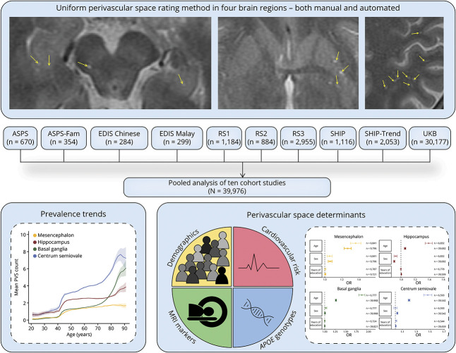
Perivascular spaces (PVS) are emerging markers of cerebral small vessel disease (CSVD), but research on their determinants has been hampered by conflicting results from small single studies using heterogeneous rating methods. In this study, we therefore aimed to identify determinants of PVS burden in a pooled analysis of multiple cohort studies using 1 harmonized PVS rating method.
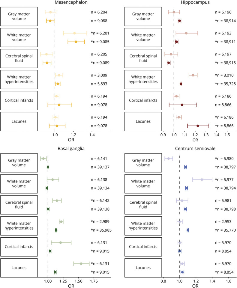
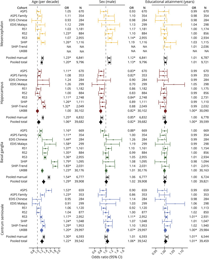
Individuals from 10 population-based cohort studies with adult participants from the Uniform Neuro-Imaging of Virchow-Robin Spaces Enlargement consortium and the UK Biobank were included. On MRI scans, we counted PVS in 4 brain regions (mesencephalon, hippocampus, basal ganglia, and centrum semiovale) according to a uniform and validated rating protocol, both manually and automated using a deep learning algorithm. As potential determinants, we considered demographics, cardiovascular risk factors, genotypes, and other imaging markers of CSVD. Negative binomial regression models were used to examine the association between these determinants and PVS counts.
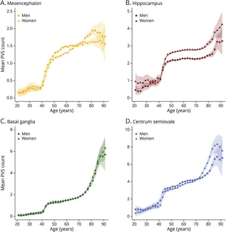
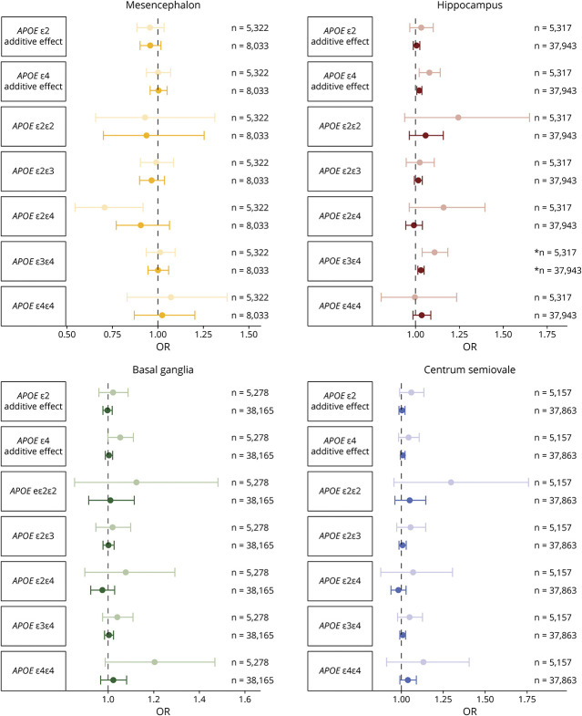
In total, 39,976 individuals were included (age range 20-96 years). The average count of PVS in the 4 regions increased from the age 20 years (0-1 PVS) to 90 years (2-7 PVS). Men had more mesencephalic PVS (OR [95% CI] = 1.13 [1.08-1.18] compared with women), but less hippocampal PVS (0.82 [0.81-0.83]). Higher blood pressure, particularly diastolic pressure, was associated with more PVS in all regions (ORs between 1.04-1.05). Hippocampal PVS showed higher counts with higher high-density lipoprotein cholesterol levels (1.02 [1.01-1.02]), glucose levels (1.02 [1.01-1.03]), and ε4-alleles (1.02 [1.01-1.04]). Furthermore, white matter hyperintensity volume and presence of lacunes were associated with PVS in multiple regions, but most strongly with the basal ganglia (1.13 [1.12-1.14] and 1.10 [1.09-1.12], respectively).
Various factors are associated with the burden of PVS, in part regionally specific, which points toward a multifactorial origin beyond what can be expected from PVS-related risk factor profiles. This study highlights the power of collaborative efforts in population neuroimaging research.
血管周围间隙(PVS)是脑小血管病(CSVD)的新兴标志物,但由于小样本单中心研究采用不同的评分方法,导致其决定因素的研究结果相互矛盾。因此,本研究旨在使用一种统一的 PVS 评分方法,对多个队列研究进行汇总分析,以确定 PVS 负荷的决定因素。
本研究纳入了来自统一神经影像学研究血管周围间隙扩大联盟和英国生物库的 10 项基于人群的队列研究中的成年人。在 MRI 扫描中,我们根据统一的验证后评分方案,对 4 个脑区(中脑、海马体、基底节和半卵圆中心)的 PVS 进行计数,同时使用深度学习算法进行手动和自动计数。我们将人口统计学因素、心血管危险因素、基因型和其他 CSVD 的影像学标志物等作为潜在的决定因素。使用负二项回归模型检验这些决定因素与 PVS 计数之间的关系。
共纳入 39976 名参与者(年龄 20-96 岁)。4 个区域的 PVS 平均计数从 20 岁(0-1 个 PVS)增加到 90 岁(2-7 个 PVS)。男性的中脑 PVS 较多(与女性相比,OR [95%CI]为 1.13 [1.08-1.18]),但海马体 PVS 较少(0.82 [0.81-0.83])。较高的血压,特别是舒张压,与所有区域的 PVS 增加有关(OR 为 1.04-1.05)。高密度脂蛋白胆固醇水平(1.02 [1.01-1.02])、血糖水平(1.02 [1.01-1.03])和 ε4 等位基因(1.02 [1.01-1.04])较高的个体,其海马体 PVS 计数较高。此外,脑白质高信号体积和腔隙存在与多个区域的 PVS 相关,但与基底节的相关性最强(分别为 1.13 [1.12-1.14]和 1.10 [1.09-1.12])。
各种因素与 PVS 负荷有关,部分与特定区域有关,这表明 PVS 的多因素起源超出了 PVS 相关危险因素谱的预期。本研究强调了在人群神经影像学研究中开展合作努力的重要性。