• 文献检索
  • 文档翻译
  • 深度研究
  • 学术资讯
  • Suppr Zotero 插件Zotero 插件
  • 邀请有礼
  • 套餐&价格
  • 历史记录
应用&插件
Suppr Zotero 插件Zotero 插件浏览器插件Mac 客户端Windows 客户端微信小程序
定价
高级版会员购买积分包购买API积分包
服务
文献检索文档翻译深度研究API 文档MCP 服务
关于我们
关于 Suppr公司介绍联系我们用户协议隐私条款
关注我们

Suppr 超能文献

核心技术专利:CN118964589B侵权必究
粤ICP备2023148730 号-1Suppr @ 2026

文献检索

告别复杂PubMed语法,用中文像聊天一样搜索,搜遍4000万医学文献。AI智能推荐,让科研检索更轻松。

立即免费搜索

文件翻译

保留排版,准确专业,支持PDF/Word/PPT等文件格式,支持 12+语言互译。

免费翻译文档

深度研究

AI帮你快速写综述,25分钟生成高质量综述,智能提取关键信息,辅助科研写作。

立即免费体验

扫描仪变异性对计算病理学中淋巴结分割的影响。

Impact of scanner variability on lymph node segmentation in computational pathology.

作者信息

Khan Amjad, Janowczyk Andrew, Müller Felix, Blank Annika, Nguyen Huu Giao, Abbet Christian, Studer Linda, Lugli Alessandro, Dawson Heather, Thiran Jean-Philippe, Zlobec Inti

机构信息

Institute of Pathology, University of Bern, Murtenstrasse 31, CH-3008 Bern, Switzerland.

Case Western Reserve University, Department of Biomedical Engineering, Cleveland, OH 44106, USA.

出版信息

J Pathol Inform. 2022 Jul 25;13:100127. doi: 10.1016/j.jpi.2022.100127. eCollection 2022.

DOI:10.1016/j.jpi.2022.100127
PMID:36268105
原文链接:https://pmc.ncbi.nlm.nih.gov/articles/PMC9577043/
Abstract

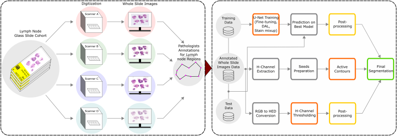
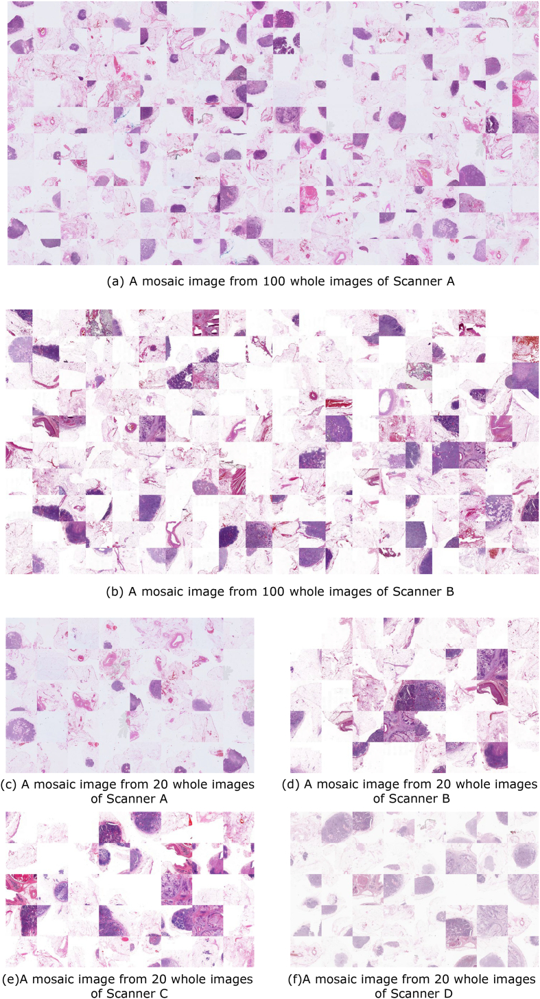
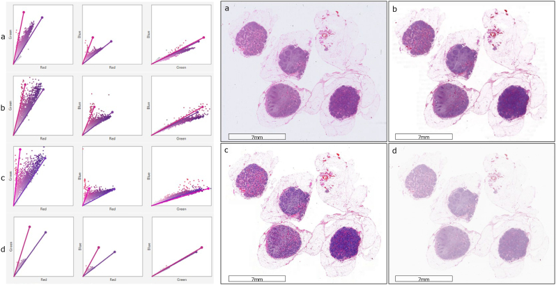
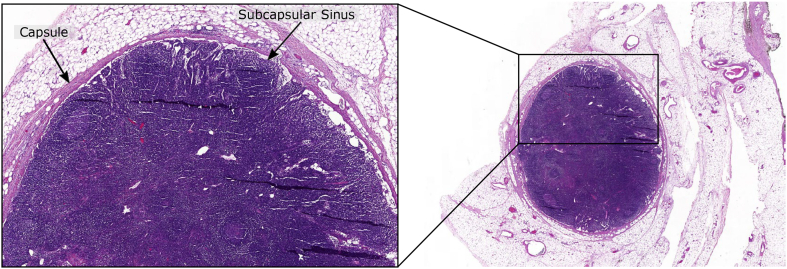
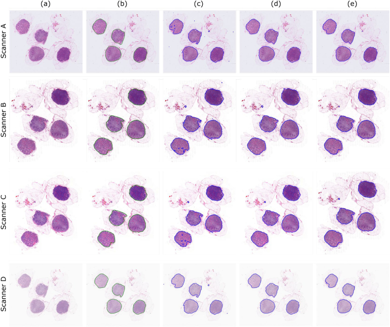
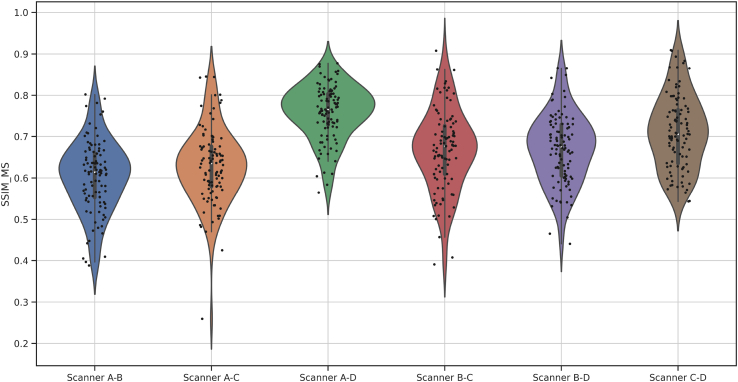
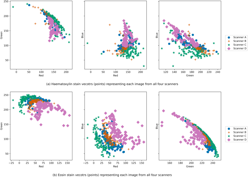
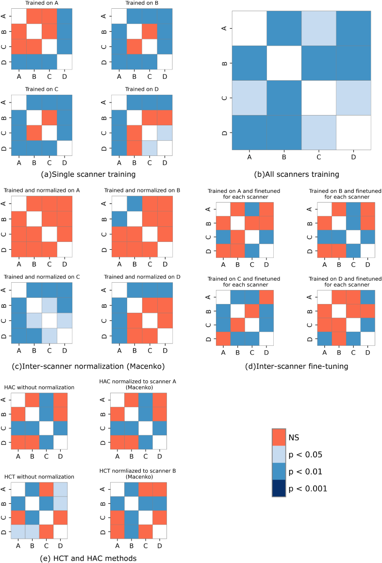
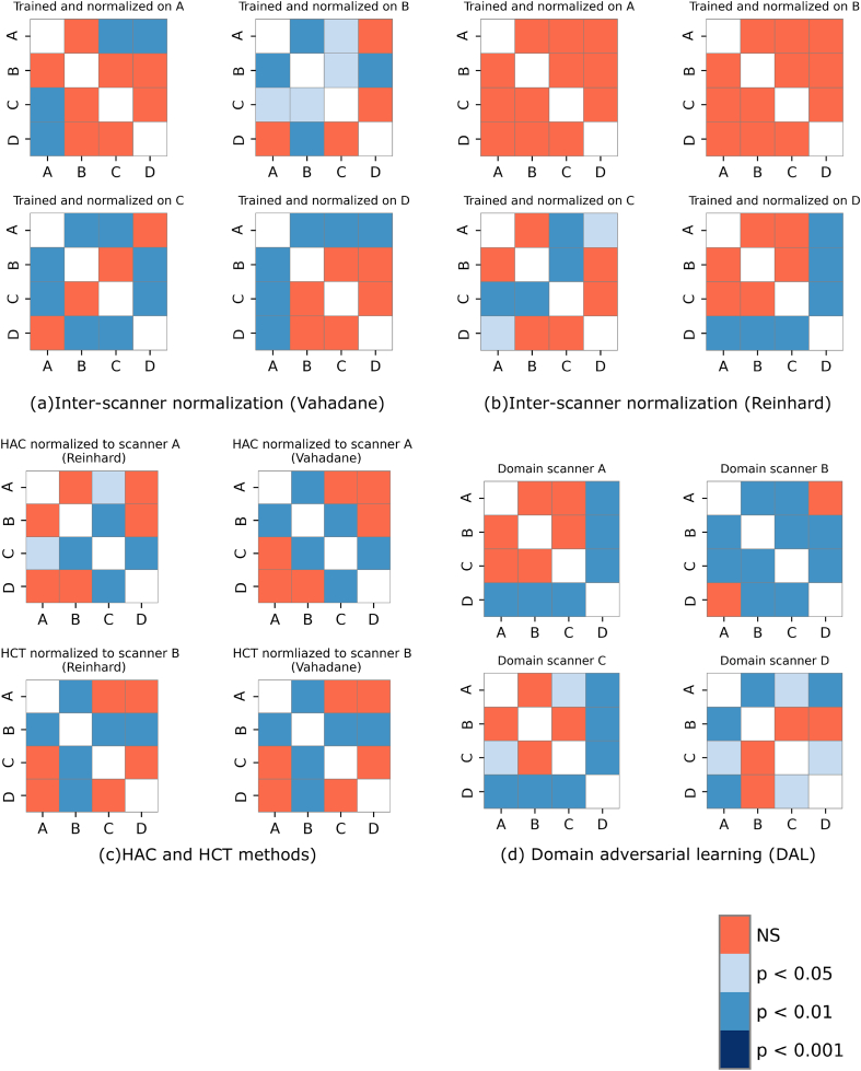
Computer-aided diagnostics in histopathology are based on the digitization of glass slides. However, heterogeneity between the images generated by different slide scanners can unfavorably affect the performance of computational algorithms. Here, we evaluate the impact of scanner variability on lymph node segmentation due to its clinical importance in colorectal cancer diagnosis. 100 slides containing 276 lymph nodes were digitized using 4 different slide scanners, and 50 of the lymph nodes containing metastatic cancer cells. These 400 scans were subsequently annotated by 2 experienced pathologists to precisely label lymph node boundary. Three different segmentation methods were then applied and compared: Hematoxylin-channel-based thresholding (HCT), Hematoxylin-based active contours (HAC), and a convolution neural network (U-Net). Evaluation of U-Net trained from both a single scanner and an ensemble of all scanners was completed. Mosaic images based on representative tiles from a scanner were used as a reference image to normalize the new data from different test scanners to evaluate the performance of a pre-trained model. Fine-tuning was carried out by using weights of a model trained on one scanner to initialize model weights for other scanners. To evaluate the domain generalization, domain adversarial learning and stain mix-up augmentation were also implemented. Results show that fine-tuning and domain adversarial learning decreased the impact of scanner variability and greatly improved segmentation across scanners. Overall, U-Net with stain mix-up (Matthews correlation coefficient (MCC) = 0.87), domain adversarial learning (MCC = 0.86), and HAC (MCC = 0.87) were shown to outperform HCT (MCC = 0.81) for segmentation of lymph nodes when compared against the ground truth. The findings of this study should be considered for future algorithms applied in diagnostic routines.

摘要

组织病理学中的计算机辅助诊断基于玻璃切片的数字化。然而,不同玻片扫描仪生成的图像之间的异质性可能会对计算算法的性能产生不利影响。在此,由于其在结直肠癌诊断中的临床重要性,我们评估了扫描仪变异性对淋巴结分割的影响。使用4种不同的玻片扫描仪对包含276个淋巴结的100张玻片进行数字化处理,其中50个淋巴结含有转移癌细胞。随后,由2名经验丰富的病理学家对这400次扫描进行注释,以精确标记淋巴结边界。然后应用并比较了三种不同的分割方法:基于苏木精通道的阈值分割(HCT)、基于苏木精的活动轮廓(HAC)和卷积神经网络(U-Net)。完成了从单个扫描仪和所有扫描仪的集合中训练U-Net的评估。基于来自扫描仪的代表性切片的拼接图像被用作参考图像,以对来自不同测试扫描仪的新数据进行归一化,从而评估预训练模型的性能。通过使用在一个扫描仪上训练的模型的权重来初始化其他扫描仪的模型权重进行微调。为了评估域泛化,还实施了域对抗学习和染色混合增强。结果表明,微调与域对抗学习降低了扫描仪变异性的影响,并大大提高了跨扫描仪的分割效果。总体而言,与真实情况相比,具有染色混合(马修斯相关系数(MCC)=0.87)、域对抗学习(MCC=0.86)和HAC(MCC=0.87)的U-Net在淋巴结分割方面表现优于HCT(MCC=0.81)。本研究的结果应被应用于未来诊断程序的算法中。

https://cdn.ncbi.nlm.nih.gov/pmc/blobs/d3df/9577043/4c594f39cc11/fx5.jpg
https://cdn.ncbi.nlm.nih.gov/pmc/blobs/d3df/9577043/7ccfde57685a/gr1.jpg
https://cdn.ncbi.nlm.nih.gov/pmc/blobs/d3df/9577043/141a5e9f4020/gr2.jpg
https://cdn.ncbi.nlm.nih.gov/pmc/blobs/d3df/9577043/c54cb4f0cb56/gr3.jpg
https://cdn.ncbi.nlm.nih.gov/pmc/blobs/d3df/9577043/b024011b0bb1/gr4.jpg
https://cdn.ncbi.nlm.nih.gov/pmc/blobs/d3df/9577043/52fbfbb0efe4/fx1.jpg
https://cdn.ncbi.nlm.nih.gov/pmc/blobs/d3df/9577043/2555e275350e/fx2.jpg
https://cdn.ncbi.nlm.nih.gov/pmc/blobs/d3df/9577043/fb3a35b5ae45/fx3.jpg
https://cdn.ncbi.nlm.nih.gov/pmc/blobs/d3df/9577043/7f7fa5175b23/fx4.jpg
https://cdn.ncbi.nlm.nih.gov/pmc/blobs/d3df/9577043/4c594f39cc11/fx5.jpg

相似文献

1
Impact of scanner variability on lymph node segmentation in computational pathology.扫描仪变异性对计算病理学中淋巴结分割的影响。
J Pathol Inform. 2022 Jul 25;13:100127. doi: 10.1016/j.jpi.2022.100127. eCollection 2022.
2
Computer-Assisted Diagnosis of Lymph Node Metastases in Colorectal Cancers Using Transfer Learning With an Ensemble Model.使用集成模型的迁移学习辅助诊断结直肠癌淋巴结转移的计算机系统。
Mod Pathol. 2023 May;36(5):100118. doi: 10.1016/j.modpat.2023.100118. Epub 2023 Feb 2.
3
Whole slide imaging (WSI) scanner differences influence optical and computed properties of digitized prostate cancer histology.全切片成像(WSI)扫描仪的差异会影响数字化前列腺癌组织学的光学和计算属性。
J Pathol Inform. 2023 Jul 4;14:100321. doi: 10.1016/j.jpi.2023.100321. eCollection 2023.
4
The Use of Deep Learning-Based Computer Diagnostic Algorithm for Detection of Lymph Node Metastases of Gastric Adenocarcinoma.基于深度学习的计算机诊断算法在检测胃腺癌淋巴结转移中的应用
Int J Surg Pathol. 2023 Sep;31(6):975-981. doi: 10.1177/10668969221113475. Epub 2022 Jul 27.
5
Improving segmentation reliability of multi-scanner brain images using a generative adversarial network.使用生成对抗网络提高多扫描仪脑图像的分割可靠性
Quant Imaging Med Surg. 2022 Mar;12(3):1775-1786. doi: 10.21037/qims-21-653.
6
Automatic segmentation of the thalamus using a massively trained 3D convolutional neural network: higher sensitivity for the detection of reduced thalamus volume by improved inter-scanner stability.使用大规模训练的 3D 卷积神经网络进行丘脑自动分割:通过提高扫描仪间稳定性来提高检测丘脑体积减小的灵敏度。
Eur Radiol. 2023 Mar;33(3):1852-1861. doi: 10.1007/s00330-022-09170-y. Epub 2022 Oct 20.
7
ARPM-net: A novel CNN-based adversarial method with Markov random field enhancement for prostate and organs at risk segmentation in pelvic CT images.ARPM-net:一种新颖的基于 CNN 的对抗性方法,结合马尔可夫随机场增强,用于骨盆 CT 图像中的前列腺和危及器官分割。
Med Phys. 2021 Jan;48(1):227-237. doi: 10.1002/mp.14580. Epub 2020 Nov 24.
8
A multiple-channel and atrous convolution network for ultrasound image segmentation.一种用于超声图像分割的多通道多孔卷积网络。
Med Phys. 2020 Dec;47(12):6270-6285. doi: 10.1002/mp.14512. Epub 2020 Oct 18.
9
Diagnostic validation of a portable whole slide imaging scanner for lymphoma diagnosis in resource-constrained setting: A cross-sectional study.资源受限环境下用于淋巴瘤诊断的便携式全切片成像扫描仪的诊断验证:一项横断面研究。
J Pathol Inform. 2023 Jan 9;14:100188. doi: 10.1016/j.jpi.2023.100188. eCollection 2023.
10
Robustness of apparent diffusion coefficient-based lymph node classification for diagnosis of prostate cancer metastasis.基于表观扩散系数的淋巴结分类对前列腺癌转移诊断的稳健性。
Eur Radiol. 2024 Jul;34(7):4504-4515. doi: 10.1007/s00330-023-10406-8. Epub 2023 Dec 15.

引用本文的文献

1
Multicenter Histology Image Integration and Multiscale Deep Learning for Machine Learning-Enabled Pediatric Sarcoma Classification.用于支持机器学习的小儿肉瘤分类的多中心组织学图像整合与多尺度深度学习
medRxiv. 2025 Jun 11:2025.06.10.25328700. doi: 10.1101/2025.06.10.25328700.
2
Rethinking model prototyping through the MedMNIST+ dataset collection.通过MedMNIST+数据集收集重新思考模型原型设计。
Sci Rep. 2025 Mar 5;15(1):7669. doi: 10.1038/s41598-025-92156-9.
3
Out-of-distribution generalization for segmentation of lymph node metastasis in breast cancer.

本文引用的文献

1
Measuring Domain Shift for Deep Learning in Histopathology.深度学习在组织病理学中的领域迁移测量。
IEEE J Biomed Health Inform. 2021 Feb;25(2):325-336. doi: 10.1109/JBHI.2020.3032060. Epub 2021 Feb 5.
2
Impact of rescanning and normalization on convolutional neural network performance in multi-center, whole-slide classification of prostate cancer.在前列腺癌的多中心全切片分类中,重新扫描和归一化对卷积神经网络性能的影响。
Sci Rep. 2020 Sep 1;10(1):14398. doi: 10.1038/s41598-020-71420-0.
3
NuClick: A deep learning framework for interactive segmentation of microscopic images.
用于乳腺癌淋巴结转移分割的分布外泛化
Sci Rep. 2025 Jan 7;15(1):1127. doi: 10.1038/s41598-024-80495-y.
4
Computational methods for metastasis detection in lymph nodes and characterization of the metastasis-free lymph node microarchitecture: A systematic-narrative hybrid review.淋巴结转移检测及无转移淋巴结微结构特征分析的计算方法:一项系统叙述性混合综述
J Pathol Inform. 2024 Feb 4;15:100367. doi: 10.1016/j.jpi.2024.100367. eCollection 2024 Dec.
5
Data-driven color augmentation for H&E stained images in computational pathology.计算病理学中用于苏木精-伊红(H&E)染色图像的数据驱动颜色增强
J Pathol Inform. 2023 Jan 3;14:100183. doi: 10.1016/j.jpi.2022.100183. eCollection 2023.
6
Digital pathology implementation in a private laboratory: The CEDAP experience.私立实验室中的数字病理学实施:CEDAP经验
J Pathol Inform. 2023 Jan 3;14:100180. doi: 10.1016/j.jpi.2022.100180. eCollection 2023.
7
Stain normalization in digital pathology: Clinical multi-center evaluation of image quality.数字病理学中的染色标准化:图像质量的临床多中心评估
J Pathol Inform. 2022 Sep 24;13:100145. doi: 10.1016/j.jpi.2022.100145. eCollection 2022.
NuClick:用于微观图像交互式分割的深度学习框架。
Med Image Anal. 2020 Oct;65:101771. doi: 10.1016/j.media.2020.101771. Epub 2020 Jul 10.
4
Whole slide images based cancer survival prediction using attention guided deep multiple instance learning networks.基于全切片图像的注意力引导深度多实例学习网络的癌症生存预测。
Med Image Anal. 2020 Oct;65:101789. doi: 10.1016/j.media.2020.101789. Epub 2020 Jul 19.
5
The advantages of the Matthews correlation coefficient (MCC) over F1 score and accuracy in binary classification evaluation.马修斯相关系数(MCC)在二分类评估中优于 F1 得分和准确率的优势。
BMC Genomics. 2020 Jan 2;21(1):6. doi: 10.1186/s12864-019-6413-7.
6
Assessing the Impact of Color Normalization in Convolutional Neural Network-Based Nuclei Segmentation Frameworks.评估颜色归一化在基于卷积神经网络的细胞核分割框架中的影响。
Front Bioeng Biotechnol. 2019 Nov 1;7:300. doi: 10.3389/fbioe.2019.00300. eCollection 2019.
7
Staining Invariant Features for Improving Generalization of Deep Convolutional Neural Networks in Computational Pathology.用于提高计算病理学中深度卷积神经网络泛化能力的染色不变特征
Front Bioeng Biotechnol. 2019 Aug 23;7:198. doi: 10.3389/fbioe.2019.00198. eCollection 2019.
8
Quantifying the effects of data augmentation and stain color normalization in convolutional neural networks for computational pathology.量化数据增强和染色颜色归一化在计算病理学卷积神经网络中的作用。
Med Image Anal. 2019 Dec;58:101544. doi: 10.1016/j.media.2019.101544. Epub 2019 Aug 21.
9
Clinical-grade computational pathology using weakly supervised deep learning on whole slide images.基于全切片图像的弱监督深度学习的临床级计算病理学。
Nat Med. 2019 Aug;25(8):1301-1309. doi: 10.1038/s41591-019-0508-1. Epub 2019 Jul 15.
10
Adaptive color deconvolution for histological WSI normalization.自适应颜色反卷积用于组织学 WSI 归一化。
Comput Methods Programs Biomed. 2019 Mar;170:107-120. doi: 10.1016/j.cmpb.2019.01.008. Epub 2019 Jan 15.