• 文献检索
  • 文档翻译
  • 深度研究
  • 学术资讯
  • Suppr Zotero 插件Zotero 插件
  • 邀请有礼
  • 套餐&价格
  • 历史记录
应用&插件
Suppr Zotero 插件Zotero 插件浏览器插件Mac 客户端Windows 客户端微信小程序
定价
高级版会员购买积分包购买API积分包
服务
文献检索文档翻译深度研究API 文档MCP 服务
关于我们
关于 Suppr公司介绍联系我们用户协议隐私条款
关注我们

Suppr 超能文献

核心技术专利:CN118964589B侵权必究
粤ICP备2023148730 号-1Suppr @ 2026

文献检索

告别复杂PubMed语法,用中文像聊天一样搜索,搜遍4000万医学文献。AI智能推荐,让科研检索更轻松。

立即免费搜索

文件翻译

保留排版,准确专业,支持PDF/Word/PPT等文件格式,支持 12+语言互译。

免费翻译文档

深度研究

AI帮你快速写综述,25分钟生成高质量综述,智能提取关键信息,辅助科研写作。

立即免费体验

CODA:用于细胞分辨率的大型组织的定量 3D 重建。

CODA: quantitative 3D reconstruction of large tissues at cellular resolution.

机构信息

Department of Chemical and Biomolecular Engineering, The Johns Hopkins University, Baltimore, MD, USA.

Department of Pathology, The Sol Goldman Pancreatic Cancer Research Center, The Johns Hopkins University School of Medicine, Baltimore, MD, USA.

出版信息

Nat Methods. 2022 Nov;19(11):1490-1499. doi: 10.1038/s41592-022-01650-9. Epub 2022 Oct 24.

DOI:10.1038/s41592-022-01650-9
PMID:36280719
原文链接:https://pmc.ncbi.nlm.nih.gov/articles/PMC10500590/
Abstract

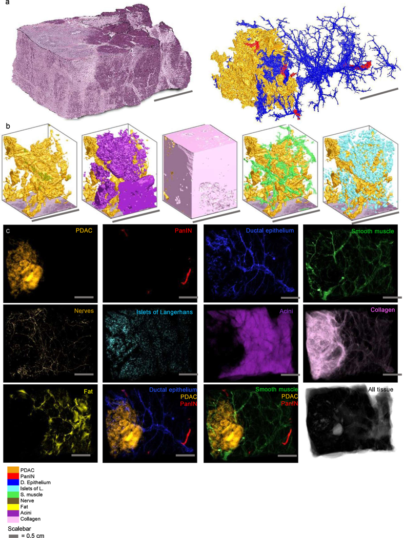
A central challenge in biology is obtaining high-content, high-resolution information while analyzing tissue samples at volumes relevant to disease progression. We address this here with CODA, a method to reconstruct exceptionally large (up to multicentimeter cubed) tissues at subcellular resolution using serially sectioned hematoxylin and eosin-stained tissue sections. Here we demonstrate CODA's ability to reconstruct three-dimensional (3D) distinct microanatomical structures in pancreas, skin, lung and liver tissues. CODA allows creation of readily quantifiable tissue volumes amenable to biological research. As a testbed, we assess the microanatomy of the human pancreas during tumorigenesis within the branching pancreatic ductal system, labeling ten distinct structures to examine heterogeneity and structural transformation during neoplastic progression. We show that pancreatic precancerous lesions develop into distinct 3D morphological phenotypes and that pancreatic cancer tends to spread far from the bulk tumor along collagen fibers that are highly aligned to the 3D curves of ductal, lobular, vascular and neural structures. Thus, CODA establishes a means to transform broadly the structural study of human diseases through exploration of exhaustively labeled 3D microarchitecture.

摘要

生物学的一个核心挑战是在分析与疾病进展相关的组织样本时,获取高含量、高分辨率的信息。我们在这里使用 CODA 解决了这个问题,这是一种使用连续切片的苏木精和伊红染色组织切片以亚细胞分辨率重建异常大(高达立方厘米级)组织的方法。在这里,我们展示了 CODA 重建胰腺、皮肤、肺和肝脏组织中三维(3D)不同微观解剖结构的能力。CODA 允许创建易于量化的组织体积,适用于生物学研究。作为一个测试平台,我们评估了分支胰腺导管系统中肿瘤发生过程中人类胰腺的微观解剖结构,标记了十个不同的结构,以检查在肿瘤进展过程中的异质性和结构转化。我们表明,胰腺癌前病变发展成独特的 3D 形态表型,并且胰腺癌往往沿着与导管、小叶、血管和神经结构的 3D 曲线高度对齐的胶原纤维远离肿瘤主体扩散。因此,CODA 通过探索详尽标记的 3D 微观结构,为广泛探索人类疾病的结构研究提供了一种手段。

https://cdn.ncbi.nlm.nih.gov/pmc/blobs/2dd4/10500590/dab291beec59/nihms-1905018-f0006.jpg
https://cdn.ncbi.nlm.nih.gov/pmc/blobs/2dd4/10500590/731aaeb5b826/nihms-1905018-f0007.jpg
https://cdn.ncbi.nlm.nih.gov/pmc/blobs/2dd4/10500590/88dd0ccf5a80/nihms-1905018-f0008.jpg
https://cdn.ncbi.nlm.nih.gov/pmc/blobs/2dd4/10500590/3cd83a473c19/nihms-1905018-f0009.jpg
https://cdn.ncbi.nlm.nih.gov/pmc/blobs/2dd4/10500590/13f5b6bf1186/nihms-1905018-f0010.jpg
https://cdn.ncbi.nlm.nih.gov/pmc/blobs/2dd4/10500590/2aaf375ea317/nihms-1905018-f0011.jpg
https://cdn.ncbi.nlm.nih.gov/pmc/blobs/2dd4/10500590/df78ad4cdc9b/nihms-1905018-f0012.jpg
https://cdn.ncbi.nlm.nih.gov/pmc/blobs/2dd4/10500590/c2515d8c94fe/nihms-1905018-f0013.jpg
https://cdn.ncbi.nlm.nih.gov/pmc/blobs/2dd4/10500590/2555fd805c2e/nihms-1905018-f0014.jpg
https://cdn.ncbi.nlm.nih.gov/pmc/blobs/2dd4/10500590/a476e57daee0/nihms-1905018-f0001.jpg
https://cdn.ncbi.nlm.nih.gov/pmc/blobs/2dd4/10500590/bfc5d924563c/nihms-1905018-f0002.jpg
https://cdn.ncbi.nlm.nih.gov/pmc/blobs/2dd4/10500590/ca41af6b4813/nihms-1905018-f0003.jpg
https://cdn.ncbi.nlm.nih.gov/pmc/blobs/2dd4/10500590/4d1f75048046/nihms-1905018-f0004.jpg
https://cdn.ncbi.nlm.nih.gov/pmc/blobs/2dd4/10500590/347c0970c6ed/nihms-1905018-f0005.jpg
https://cdn.ncbi.nlm.nih.gov/pmc/blobs/2dd4/10500590/dab291beec59/nihms-1905018-f0006.jpg

相似文献

1
CODA: quantitative 3D reconstruction of large tissues at cellular resolution.CODA:用于细胞分辨率的大型组织的定量 3D 重建。
Nat Methods. 2022 Nov;19(11):1490-1499. doi: 10.1038/s41592-022-01650-9. Epub 2022 Oct 24.
2
Tissue clearing and 3D reconstruction of digitized, serially sectioned slides provide novel insights into pancreatic cancer.数字化、连续切片的组织透明化和 3D 重建为胰腺癌研究提供了新的视角。
Med. 2023 Feb 10;4(2):75-91. doi: 10.1016/j.medj.2022.11.009. Epub 2023 Jan 24.
3
A "Clearer" View of Pancreatic Pathology: A Review of Tissue Clearing and Advanced Microscopy Techniques.更清晰的胰腺病理学视野:组织透明化和高级显微镜技术综述。
Adv Anat Pathol. 2019 Jan;26(1):31-39. doi: 10.1097/PAP.0000000000000215.
4
A Study of the Growth Patterns of Breast Carcinoma Using 3D Reconstruction: A Pilot Study.使用三维重建技术对乳腺癌生长模式的研究:一项初步研究。
Breast J. 2017 Jan;23(1):83-89. doi: 10.1111/tbj.12688. Epub 2016 Nov 17.
5
Robust 3D image reconstruction of pancreatic cancer tumors from histopathological images with different stains and its quantitative performance evaluation.利用不同染色的组织病理学图像对胰腺癌肿瘤进行稳健的三维图像重建及其定量性能评估。
Int J Comput Assist Radiol Surg. 2019 Dec;14(12):2047-2055. doi: 10.1007/s11548-019-02019-8. Epub 2019 Jul 2.
6
Three-dimensional assessments are necessary to determine the true, spatially-resolved composition of tissues.三维评估对于确定组织的真实、空间分辨组成是必要的。
bioRxiv. 2024 Mar 28:2023.12.04.569986. doi: 10.1101/2023.12.04.569986.
7
Quantitative assessment of pancreatic cancer precursor lesions in IHC-stained tissue with a tissue image analysis platform.使用组织图像分析平台对免疫组化染色组织中的胰腺癌前体病变进行定量评估。
Lab Invest. 2016 Dec;96(12):1327-1336. doi: 10.1038/labinvest.2016.111. Epub 2016 Oct 24.
8
A role of three-dimensional (3D) reconstruction in the classification of lung adenocarcinoma.三维(3D)重建在肺腺癌分类中的作用。
Stud Health Technol Inform. 2012;179:250-6.
9
Three-dimensional analysis of human pancreatic cancer specimens by phase-contrast based X-ray tomography - the next dimension of diagnosis.基于相衬 X 射线断层摄影术的人类胰腺癌标本的三维分析——诊断的下一个维度。
Cancer Imaging. 2023 May 2;23(1):43. doi: 10.1186/s40644-023-00559-6.
10
Mesoscopic 3D imaging of pancreatic cancer and Langerhans islets based on tissue autofluorescence.基于组织自发荧光的胰腺癌和胰岛的介观 3D 成像。
Sci Rep. 2020 Oct 26;10(1):18246. doi: 10.1038/s41598-020-74616-6.

引用本文的文献

1
Spatial Multiplexing and Omics.空间复用与组学
Nat Rev Methods Primers. 2024;4(1). doi: 10.1038/s43586-024-00330-6. Epub 2024 Aug 1.
2
Generating 2.5D pathology for enhanced viewing and AI diagnosis.生成2.5D病理学图像以增强观察效果和人工智能诊断。
J Pathol Inform. 2025 Jul 18;18:100463. doi: 10.1016/j.jpi.2025.100463. eCollection 2025 Aug.
3
Neoadjuvant immunotherapy promotes the formation of mature tertiary lymphoid structures in a remodeled pancreatic tumor microenvironment.新辅助免疫疗法可促进重塑的胰腺肿瘤微环境中成熟三级淋巴结构的形成。

本文引用的文献

1
Prostate Cancer Risk Stratification via Nondestructive 3D Pathology with Deep Learning-Assisted Gland Analysis.基于深度学习辅助腺体分析的非破坏性 3D 病理学前列腺癌风险分层。
Cancer Res. 2022 Jan 15;82(2):334-345. doi: 10.1158/0008-5472.CAN-21-2843. Epub 2021 Dec 1.
2
Characterization of tumor-associated macrophages in prostate cancer transgenic mouse models.前列腺癌转基因小鼠模型中肿瘤相关巨噬细胞的特征。
Prostate. 2021 Jul;81(10):629-647. doi: 10.1002/pros.24139. Epub 2021 May 5.
3
Harnessing non-destructive 3D pathology.利用非破坏性三维病理学
Cancer Immunol Res. 2025 Aug 15. doi: 10.1158/2326-6066.CIR-25-0387.
4
Deep-learning triage of 3D pathology datasets for comprehensive and efficient pathologist assessments.用于全面高效的病理学家评估的3D病理数据集的深度学习分类
bioRxiv. 2025 Jul 22:2025.07.20.665804. doi: 10.1101/2025.07.20.665804.
5
COEXIST: Coordinated single-cell integration of serial multiplexed tissue images.COEXIST:连续多重组织图像的协调单细胞整合
PLoS Comput Biol. 2025 Aug 5;21(8):e1013325. doi: 10.1371/journal.pcbi.1013325. eCollection 2025 Aug.
6
PIVOT: an open-source tool for multi-omic spatial data registration.PIVOT:一种用于多组学空间数据配准的开源工具。
bioRxiv. 2025 Jun 8:2025.06.08.658506. doi: 10.1101/2025.06.08.658506.
7
Differential cell signaling testing for cell-cell communication inference from single-cell data by dominoSignal.通过dominoSignal从单细胞数据进行细胞间通讯推断的差异细胞信号测试。
bioRxiv. 2025 May 3:2025.05.02.651747. doi: 10.1101/2025.05.02.651747.
8
Three-dimensional topological defects and quasi-long-range order in biological liquid crystals.生物液晶中的三维拓扑缺陷与准长程有序
bioRxiv. 2025 May 10:2025.04.14.648711. doi: 10.1101/2025.04.14.648711.
9
Informatics at the Frontier of Cancer Research.癌症研究前沿的信息学
Cancer Res. 2025 Aug 15;85(16):2967-2986. doi: 10.1158/0008-5472.CAN-24-2829.
10
Spatial proteomics and transcriptomics reveal early immune cell organization in pancreatic intraepithelial neoplasia.空间蛋白质组学和转录组学揭示胰腺上皮内瘤变中早期免疫细胞组织。
JCI Insight. 2025 Jun 26;10(15). doi: 10.1172/jci.insight.191595. eCollection 2025 Aug 8.
Nat Biomed Eng. 2021 Mar;5(3):203-218. doi: 10.1038/s41551-020-00681-x. Epub 2021 Feb 15.
4
Cancer Statistics, 2021.癌症统计数据,2021.
CA Cancer J Clin. 2021 Jan;71(1):7-33. doi: 10.3322/caac.21654. Epub 2021 Jan 12.
5
VISTA: VIsual Semantic Tissue Analysis for pancreatic disease quantification in murine cohorts.VISTA:用于鼠队列中胰腺疾病定量的视觉语义组织分析。
Sci Rep. 2020 Dec 1;10(1):20904. doi: 10.1038/s41598-020-78061-3.
6
Mesoscopic 3D imaging of pancreatic cancer and Langerhans islets based on tissue autofluorescence.基于组织自发荧光的胰腺癌和胰岛的介观 3D 成像。
Sci Rep. 2020 Oct 26;10(1):18246. doi: 10.1038/s41598-020-74616-6.
7
Histological 3D reconstruction and in vivo lineage tracing of the human endometrium.人类子宫内膜的组织学 3D 重建与体内谱系追踪。
J Pathol. 2020 Aug;251(4):440-451. doi: 10.1002/path.5478. Epub 2020 Jun 30.
8
Versatile whole-organ/body staining and imaging based on electrolyte-gel properties of biological tissues.基于生物组织电解质凝胶特性的多功能整体器官/组织染色和成像。
Nat Commun. 2020 Apr 27;11(1):1982. doi: 10.1038/s41467-020-15906-5.
9
Cellular and Molecular Probing of Intact Human Organs.完整人体器官的细胞和分子探测。
Cell. 2020 Feb 20;180(4):796-812.e19. doi: 10.1016/j.cell.2020.01.030. Epub 2020 Feb 13.
10
Three-dimensional visualization of cleared human pancreas cancer reveals that sustained epithelial-to-mesenchymal transition is not required for venous invasion.人类胰腺癌Cleared 组织的三维可视化显示,持续的上皮间质转化对于静脉侵犯并非必需。
Mod Pathol. 2020 Apr;33(4):639-647. doi: 10.1038/s41379-019-0409-3. Epub 2019 Nov 7.