Adl Amini Dominik, Moser Manuel, Oezel Lisa, Shue Jennifer, Pumberger Matthias, Sama Andrew A, Cammisa Frank P, Girardi Federico P, Hughes Alexander P
Spine Care Institute, Hospital for Special Surgery, New York, NY, USA.
Department of Orthopedic Surgery and Traumatology, Charité University Hospital Berlin, Berlin, Germany.
J Spine Surg. 2022 Sep;8(3):323-332. doi: 10.21037/jss-22-17.
Compare fusion at two independent timepoints (early and late) between 3D-printed titanium (Ti) and polyetheretherketone (PEEK) cages in patients undergoing standalone lateral lumbar interbody fusion (SA-LLIF). We hypothesized that 3D-printed Ti cages show higher fusion rates at an early timepoint compared to PEEK.
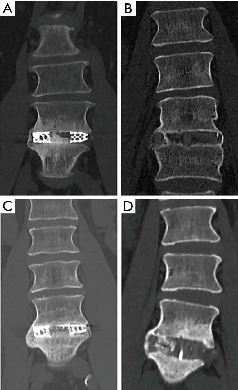
A retrospective study of patients undergoing SA-LLIF with 3D-printed Ti cages and PEEK cages between 11/2016 and 01/2020 at a single academic institution was done. Fusion was assessed for each treated level using multiplanar reconstructed computed tomography (CT) scans. Presence of fully bridged interbody trabecular bone or continuous bone centered in the cage was considered as fusion.
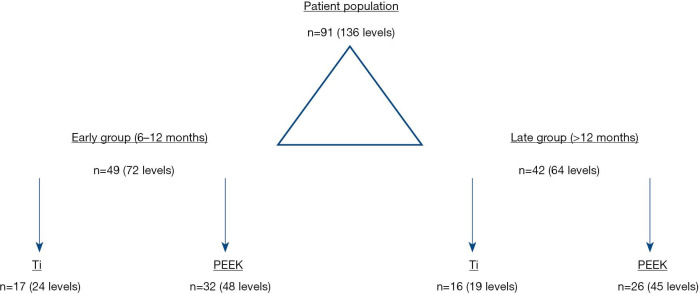
In total, 91 patients (136 levels) were included in the final analysis, 49 patients (72 levels) in the early group and 42 patients (64 levels) in the late group. CT scans were performed on average 8.2±1.8 months postoperatively for the early group and 18.9±7.7 months for the late group. In the early group, fusion was significantly higher for 3D-printed Ti cages compared to PEEK cages (95.8% versus 62.5%; P=0.002), whereas in the late group no significant difference was seen (94.7% versus 80.0%; P=0.258).
In SA-LLIF, porous 3D-printed Ti cages showed significantly higher fusion rates at an early timepoint compared to PEEK. However, the difference in fusion rates between 3D-printed Ti cages and PEEK cages was found not to be significantly different at a later timepoint in another patient group. This might support the assumption that 3D-printed Ti cages with a porous architecture are more osteoconductive compared to PEEK and tend to fuse earlier.
比较接受单纯外侧腰椎椎间融合术(SA - LLIF)的患者中,3D打印钛(Ti)笼和聚醚醚酮(PEEK)笼在两个独立时间点(早期和晚期)的融合情况。我们假设,与PEEK相比,3D打印Ti笼在早期显示出更高的融合率。
对2016年11月至2020年1月期间在单一学术机构接受SA - LLIF并使用3D打印Ti笼和PEEK笼的患者进行回顾性研究。使用多平面重建计算机断层扫描(CT)对每个治疗节段的融合情况进行评估。椎间小梁骨完全桥接或笼中心连续骨的存在被视为融合。
最终分析共纳入91例患者(136个节段),早期组49例患者(72个节段),晚期组42例患者(64个节段)。早期组术后平均8.2±1.8个月进行CT扫描,晚期组为18.9±7.7个月。在早期组中,3D打印Ti笼的融合率显著高于PEEK笼(95.8%对62.5%;P = 0.002),而在晚期组中未观察到显著差异(94.7%对80.0%;P = 0.258)。
在SA - LLIF中,与PEEK相比,多孔3D打印Ti笼在早期显示出显著更高的融合率。然而,在另一患者组的后期时间点,发现3D打印Ti笼和PEEK笼之间的融合率差异无统计学意义。这可能支持以下假设,即具有多孔结构的3D打印Ti笼比PEEK具有更高的骨传导性,并且倾向于更早融合。