Department of Nuclear Engineering, University of California, Berkeley, CA, USA.
Department of Radiology and Biomedical Imaging, University of California, San Francisco, CA, USA.
Sci Rep. 2022 Oct 26;12(1):17934. doi: 10.1038/s41598-022-22664-5.
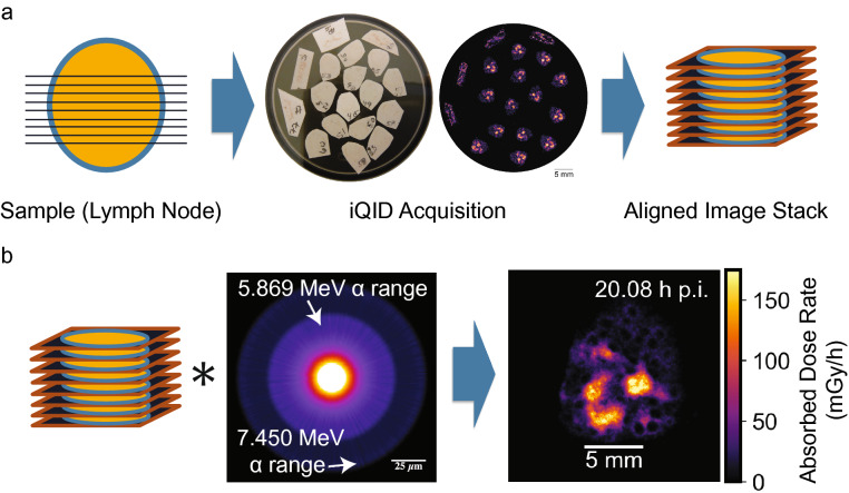
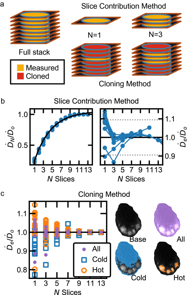
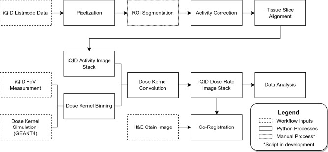
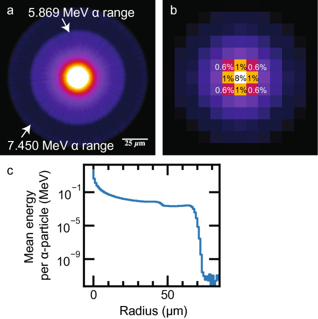
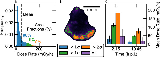
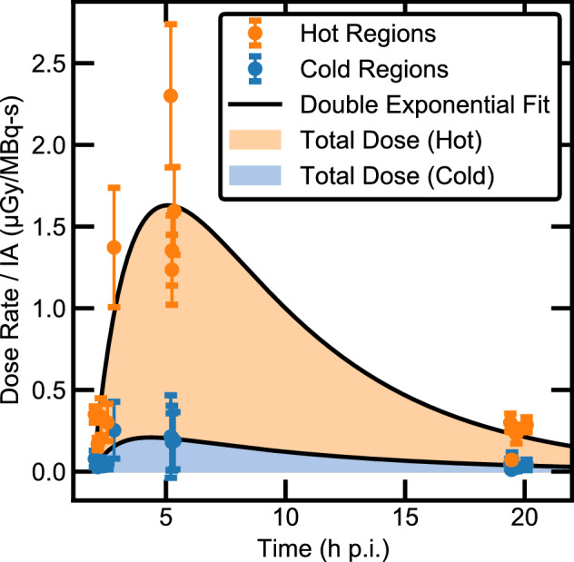
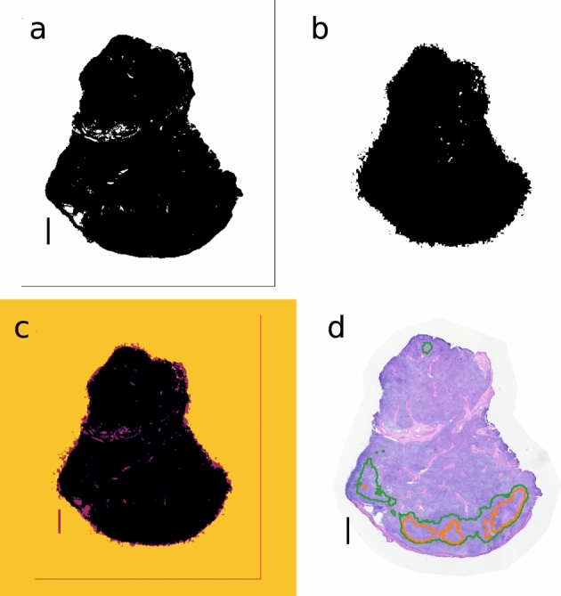
Targeted radiopharmaceutical therapy with alpha-particle emitters (αRPT) is advantageous in cancer treatment because the short range and high local energy deposition of alpha particles enable precise radiation delivery and efficient tumor cell killing. However, these properties create sub-organ dose deposition effects that are not easily characterized by direct gamma-ray imaging (PET or SPECT). We present a computational procedure to determine the spatial distribution of absorbed dose from alpha-emitting radionuclides in tissues using digital autoradiography activity images from an ionizing-radiation quantum imaging detector (iQID). Data from At-radioimmunotherapy studies for allogeneic hematopoietic cell transplantation in a canine model were used to develop these methods. Nine healthy canines were treated with 16.9-30.9 MBq At/mg monoclonal antibodies (mAb). Lymph node biopsies from early (2-5 h) and late (19-20 h) time points (16 total) were obtained, with 10-20 consecutive 12-µm cryosections extracted from each and imaged with an iQID device. iQID spatial activity images were registered within a 3D volume for dose-point-kernel convolution, producing dose-rate maps. The accumulated absorbed doses for high- and low-rate regions were 9 ± 4 Gy and 1.2 ± 0.8 Gy from separate dose-rate curves, respectively. We further assess uptake uniformity, co-registration with histological pathology, and requisite slice numbers to improve microscale characterization of absorbed dose inhomogeneities in αRPT.
利用发射阿尔法粒子的靶向放射性药物疗法(αRPT)在癌症治疗中具有优势,因为阿尔法粒子的短射程和高局部能量沉积使精确的放射治疗和高效的肿瘤细胞杀伤成为可能。然而,这些特性会产生亚器官剂量沉积效应,这些效应不容易通过直接伽马射线成像(PET 或 SPECT)来表征。我们提出了一种计算程序,使用来自离子辐射量子成像探测器(iQID)的数字放射自显影活性图像来确定组织中发射阿尔法粒子的放射性核素的吸收剂量的空间分布。这些方法是使用同种异体造血细胞移植犬模型的放射免疫治疗研究的数据开发的。将 16.9-30.9 MBq At/mg 单克隆抗体(mAb)用于 9 只健康犬的治疗。在早期(2-5 小时)和晚期(19-20 小时)时间点(共 16 个)获得淋巴结活检,从每个活检中提取 10-20 个连续的 12-µm 冷冻切片,并使用 iQID 设备进行成像。iQID 空间活性图像在 3D 体积内进行注册,以进行剂量点核函数卷积,生成剂量率图。从单独的剂量率曲线中,高和低剂量率区域的累积吸收剂量分别为 9±4 Gy 和 1.2±0.8 Gy。我们进一步评估了吸收率均匀性、与组织病理学的配准以及所需的切片数量,以改善 αRPT 中吸收剂量不均匀性的微观特征。