Clinical Physiology, Department of Clinical Sciences Lund, Lund University, Skane University Hospital, Lund, Sweden.
Wallenberg Centre for Molecular Medicine, Lund University, Lund, Sweden.
Pediatr Cardiol. 2023 Aug;44(6):1311-1318. doi: 10.1007/s00246-022-03038-0. Epub 2022 Nov 5.
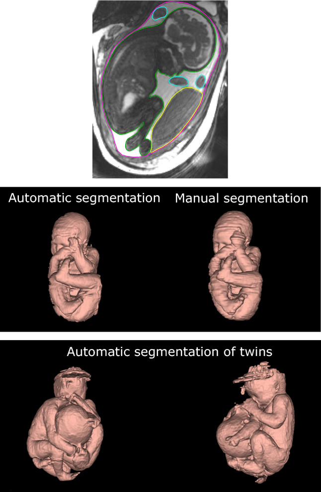
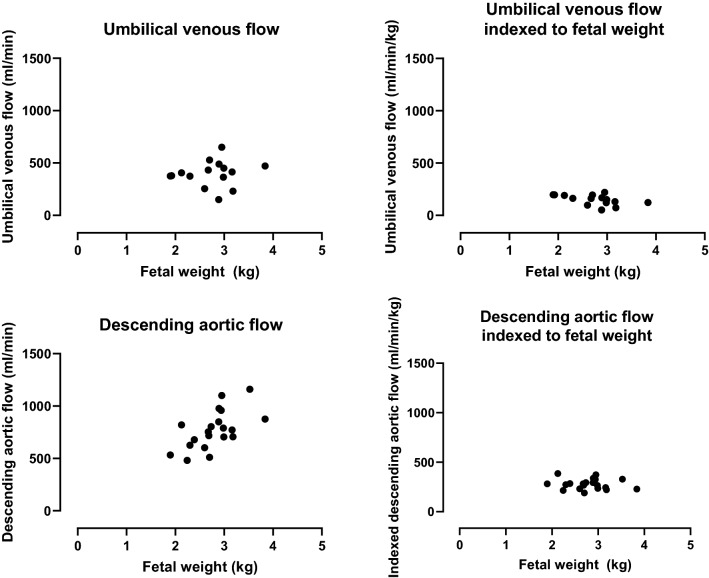
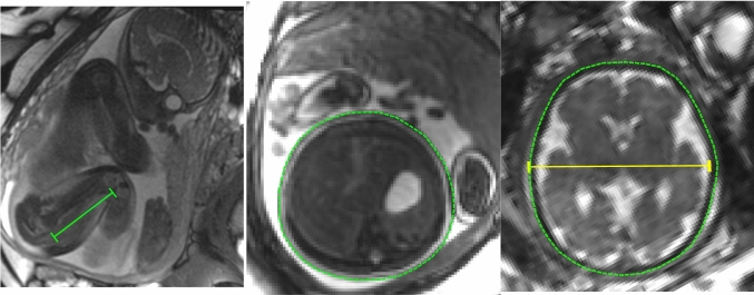
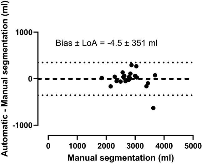
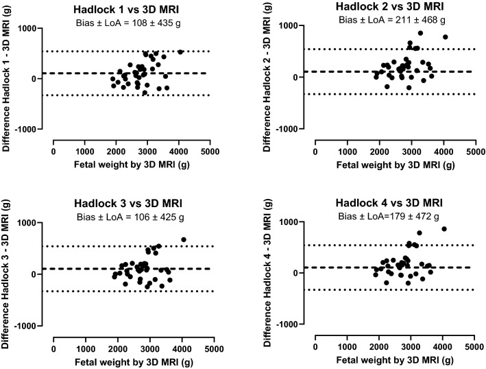
Magnetic resonance imaging (MRI) provides images for estimating fetal volume and weight, but manual delineations are time consuming. The aims were to (1) validate an algorithm to automatically quantify fetal volume by MRI; (2) compare fetal weight by Hadlock's formulas to that of MRI; and (3) quantify fetal blood flow and index flow to fetal weight by MRI. Forty-two fetuses at 36 (29-39) weeks gestation underwent MRI. A neural network was trained to segment the fetus, with 20 datasets for training and validation, and 22 for testing. Hadlock's formulas 1-4 with biometric parameters from MRI were compared with weight by MRI. Blood flow was measured using phase-contrast MRI and indexed to fetal weight. Bland-Altman analysis assessed the agreement between automatic and manual fetal segmentation and the agreement between Hadlock's formulas and fetal segmentation for fetal weight. Bias and 95% limits of agreement were for automatic versus manual measurements 4.5 ± 351 ml (0.01% ± 11%), and for Hadlock 1-4 vs MRI 108 ± 435 g (3% ± 14%), 211 ± 468 g (7% ± 15%), 106 ± 425 g (4% ± 14%), and 179 ± 472 g (6% ± 15%), respectively. Umbilical venous flow was 406 (range 151-650) ml/min (indexed 162 (range 52-220) ml/min/kg), and descending aortic flow was 763 (range 481-1160) ml/min (indexed 276 (range 189-386) ml/min/kg). The automatic method showed good agreement with manual measurements and saves considerable analysis time. Hadlock 1-4 generally agree with MRI. This study also illustrates the confounding effects of fetal weight on absolute blood flow, and emphasizes the benefit of indexed measurements for physiological assessment.
磁共振成像(MRI)可用于估计胎儿体积和体重,但手动勾画耗时。目的是:(1)验证一种自动定量 MRI 胎儿体积的算法;(2)比较 Hadlock 公式和 MRI 测量的胎儿体重;(3)定量 MRI 测量胎儿血流和指数血流与胎儿体重的关系。42 例 36 周(29-39 周)胎儿行 MRI。使用神经网络对胎儿进行分割,20 个数据集用于训练和验证,22 个数据集用于测试。比较了 MRI 生物参数的 Hadlock 公式 1-4 与 MRI 测量的体重。使用相位对比 MRI 测量血流,并与胎儿体重指数化。 Bland-Altman 分析评估了自动与手动胎儿分割之间的一致性,以及 Hadlock 公式和 MRI 胎儿体重分割之间的一致性。自动与手动测量的偏差和 95%一致性界限分别为 4.5 ± 351ml(0.01% ± 11%)和 Hadlock 1-4 与 MRI 分别为 108 ± 435g(3% ± 14%)、211 ± 468g(7% ± 15%)、106 ± 425g(4% ± 14%)和 179 ± 472g(6% ± 15%)。脐静脉血流为 406(151-650)ml/min(指数为 162(52-220)ml/min/kg),降主动脉血流为 763(481-1160)ml/min(指数为 276(189-386)ml/min/kg)。自动方法与手动测量具有良好的一致性,并节省了大量分析时间。Hadlock 1-4 通常与 MRI 一致。本研究还说明了胎儿体重对绝对血流的混杂影响,并强调了指数测量对生理评估的益处。