Indugu N, Hennessy M, Kaplan-Shabtai V S, de Assis Lage C F, Räisänen S E, Melgar A, Nedelkov K, Chen X, Oh J, Vecchiarelli B, Bender J S, Hristov A N, Pitta D W
Department of Clinical Studies, University of Pennsylvania, School of Veterinary Medicine, New Bolton Center, Kennett Square 19348.
Department of Animal Science, The Pennsylvania State University, University Park 16802.
JDS Commun. 2021 Oct 9;2(6):329-333. doi: 10.3168/jdsc.2021-0094. eCollection 2021 Nov.
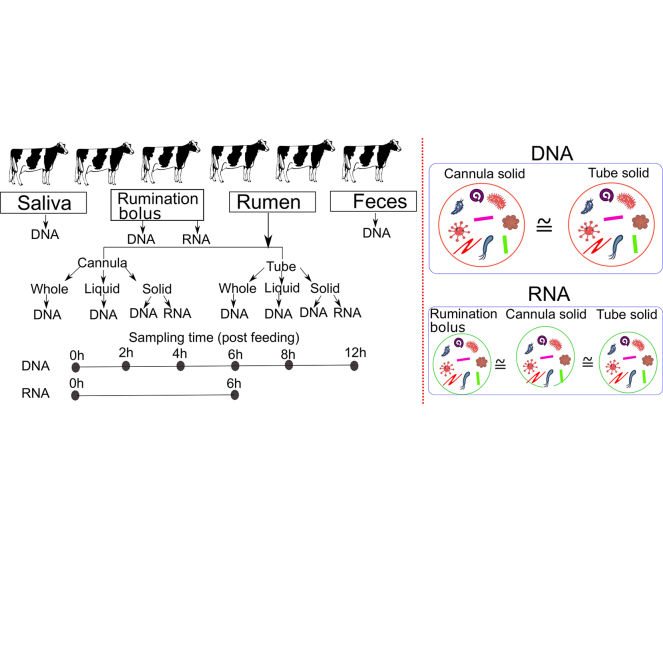
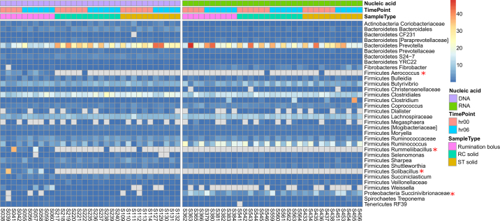
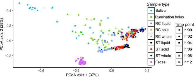
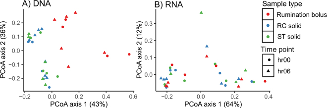
Rumen microbes play an important role in the conversion of indigestible plant material to energy and protein in dairy cows. Sampling for ruminal contents via cannula is considered the gold standard technique for microbial analysis, but the technique requires ruminally cannulated animals and specialized animal facilities. The purpose of this study was to determine whether other sampling methods and locations along the digestive tract may serve as noninvasive proxies to the cannula method for microbial analysis. Six ruminally cannulated lactating Holstein dairy cows were adapted to a standard total mixed ration for 2 wk and sampled during the third week. Sampling locations and methods included salivary content, rumination bolus (regurgitated digesta collected from the cow's mouth), feces, and rumen contents via stomach tube and cannula. Stomach tube and cannula samples differ in proportions of solid and liquid material and were therefore separated into whole (as collected), liquid, and solid fractions. Samples were collected at 0 (before feeding), 2, 4, 6, 8, and 12 h after feeding over 2 d. All samples were extracted for total genomic DNA and selected samples for metabolically active DNA (RNA), PCR-amplified for the V1-V2 region of the 16S rRNA bacterial gene, and analyzed for bacterial diversity using the QIIME2 pipeline followed by statistical analysis in R (https://www.R-project.org/). In DNA-based analysis, at the community level, saliva, rumination bolus, and fecal samples clustered in separate groups, whereas all fractions of stomach tube and cannula samples clustered together, indicating that microbial communities of stomach tube and cannula samples were homogeneous. Rumination bolus samples at 6, 8, and 12 h after feeding clustered with stomach tube and cannula samples, indicating that rumination bolus samples may be an alternative for cannula samples; however, time of sampling is critical for sampling of bolus digesta. Results of the RNA-based analysis of rumination bolus samples and solid samples from cannula and stomach tube at 0 and 6 h after feeding were similar. We concluded that the solid fraction of samples obtained via the stomach tube method may serve as a proxy for the solid fraction of whole ruminal contents obtained via cannula for DNA-based microbial investigations. Both rumination bolus and stomach tube solid samples may serve as proxies for cannula solid samples for RNA-based microbial analysis.
瘤胃微生物在奶牛将难以消化的植物性物质转化为能量和蛋白质的过程中发挥着重要作用。通过套管采集瘤胃内容物样本被认为是微生物分析的金标准技术,但该技术需要对动物进行瘤胃插管,且需要专门的动物设施。本研究的目的是确定消化道其他采样方法和部位是否可作为套管法微生物分析的非侵入性替代方法。六头装有瘤胃套管的泌乳荷斯坦奶牛适应标准全混合日粮2周,并在第三周进行采样。采样部位和方法包括唾液、反刍食团(从牛口腔收集的反刍消化物)、粪便,以及通过胃管和套管采集的瘤胃内容物。胃管和套管样本在固体和液体物质比例上存在差异,因此被分为整体(采集时)、液体和固体部分。在2天内,于喂食前(0小时)、喂食后2、4、6、8和12小时采集样本。所有样本均提取总基因组DNA,部分样本提取代谢活性DNA(RNA),对16S rRNA细菌基因的V1-V2区域进行PCR扩增,并使用QIIME2流程分析细菌多样性,随后在R(https://www.R-project.org/)中进行统计分析。在基于DNA的分析中,在群落水平上,唾液、反刍食团和粪便样本聚为不同组,而胃管和套管样本的所有部分聚在一起,表明胃管和套管样本的微生物群落是同质的。喂食后6、8和12小时的反刍食团样本与胃管和套管样本聚在一起,表明反刍食团样本可能是套管样本的替代选择;然而,采样时间对食团消化物采样至关重要。喂食后0和6小时反刍食团样本以及套管和胃管固体样本基于RNA分析的结果相似。我们得出结论,通过胃管法获得的样本固体部分可作为基于DNA的微生物研究中通过套管获得的整个瘤胃内容物固体部分的替代物。反刍食团和胃管固体样本均可作为基于RNA的微生物分析中套管固体样本的替代物。