Senol Aysegul Dilsizoglu, Pinto Giulia, Beau Maxime, Guillemot Vincent, Dupree Jeffrey L, Stadelmann Christine, Ranft Jonas, Lubetzki Catherine, Davenne Marc
Sorbonne University, Paris Brain Institute-ICM, Inserm, CNRS, Pitié-Salpêtrière Hospital, Paris, France.
Institut de Biologie de l'École Normale Supérieure (IBENS), École Normale Supérieure, CNRS, Inserm, PSL Research University, Paris, France.
Brain Commun. 2022 Nov 4;4(6):fcac284. doi: 10.1093/braincomms/fcac284. eCollection 2022.
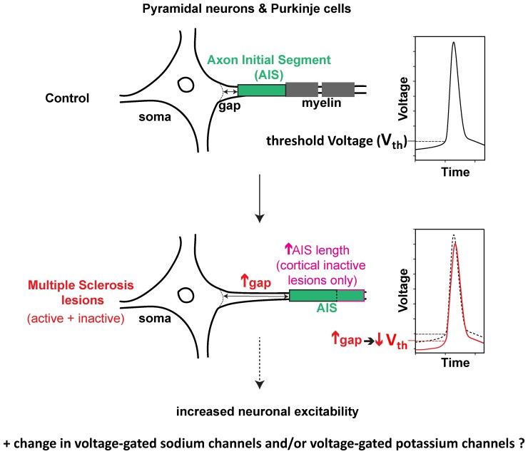
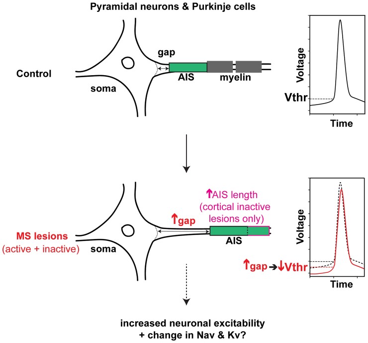
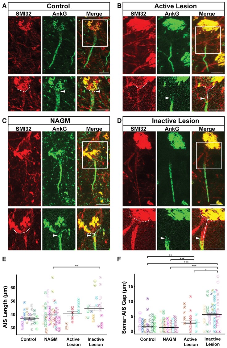
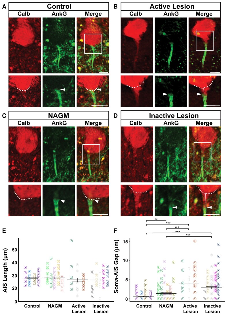
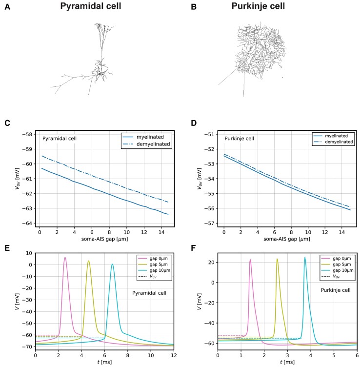
Grey matter damage has been established as a key contributor to disability progression in multiple sclerosis. Aside from neuronal loss and axonal transections, which predominate in cortical demyelinated lesions, synaptic alterations have been detected in both demyelinated plaques and normal-appearing grey matter, resulting in functional neuronal damage. The axon initial segment is a key element of neuronal function, responsible for action potential initiation and maintenance of neuronal polarity. Despite several reports of profound axon initial segment alterations in different pathological models, among which experimental auto-immune encephalomyelitis, whether the axon initial segment is affected in multiple sclerosis is still unknown. Using immunohistochemistry, we analysed axon initial segments from control and multiple sclerosis tissue, focusing on layer 5/6 pyramidal neurons in the neocortex and Purkinje cells in the cerebellum and performed analysis on the parameters known to control neuronal excitability, i.e. axon initial segment length and position. We found that the axon initial segment length was increased only in pyramidal neurons of inactive demyelinated lesions, compared with normal appearing grey matter tissue. In contrast, in both cell types, the axon initial segment position was altered, with an increased soma-axon initial segment gap, in both active and inactive demyelinated lesions. In addition, using a computational model, we show that this increased gap between soma and axon initial segment might increase neuronal excitability. Taken together, these results show, for the first time, changes of axon initial segments in multiple sclerosis, in active as well as inactive grey matter lesions in both neocortex and cerebellum, which might alter neuronal function.
灰质损伤已被确认为多发性硬化症残疾进展的关键因素。除了在皮质脱髓鞘病变中占主导地位的神经元丢失和轴突横断外,在脱髓鞘斑块和外观正常的灰质中均检测到突触改变,导致功能性神经元损伤。轴突起始段是神经元功能的关键要素,负责动作电位的起始和神经元极性的维持。尽管有几份报告称在不同病理模型(包括实验性自身免疫性脑脊髓炎)中轴突起始段有深刻改变,但轴突起始段在多发性硬化症中是否受影响仍不清楚。我们使用免疫组织化学方法,分析了对照组织和多发性硬化症组织中的轴突起始段,重点关注新皮质第5/6层锥体神经元和小脑中的浦肯野细胞,并对已知控制神经元兴奋性的参数(即轴突起始段长度和位置)进行了分析。我们发现,与外观正常的灰质组织相比,仅在非活动性脱髓鞘病变的锥体神经元中轴突起始段长度增加。相比之下,在这两种细胞类型中,无论是活动性还是非活动性脱髓鞘病变,轴突起始段位置均发生改变,胞体-轴突起始段间隙增大。此外,我们使用计算模型表明,胞体与轴突起始段之间这种增大的间隙可能会增加神经元兴奋性。综上所述,这些结果首次表明,在多发性硬化症中,新皮质和小脑中的活动性和非活动性灰质病变中的轴突起始段均发生了变化,这可能会改变神经元功能。