Neurobiology Laboratory, College of Veterinary Medicine, China Agricultural University, Haidian, Beijing 100193, China.
Key Laboratory of Precision Nutrition and Food Quality, Key Laboratory of Functional Dairy, Ministry of Education, Beijing Laboratory of Food Quality and Safety, Department of Nutrition and Health, China Agricultural University, Beijing 100083, China.
Cells. 2022 Nov 28;11(23):3808. doi: 10.3390/cells11233808.
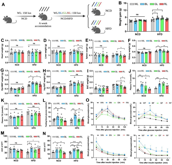
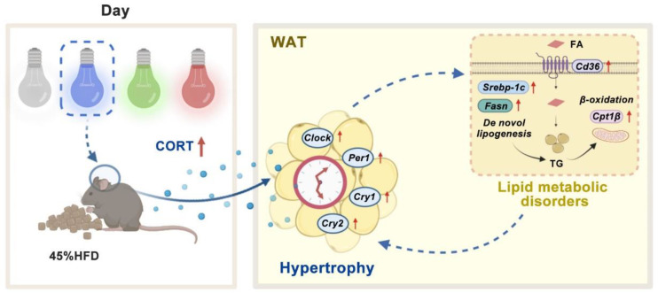
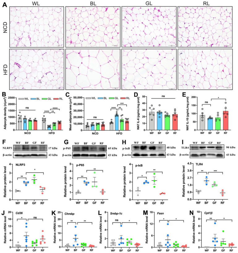
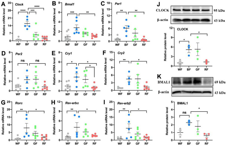
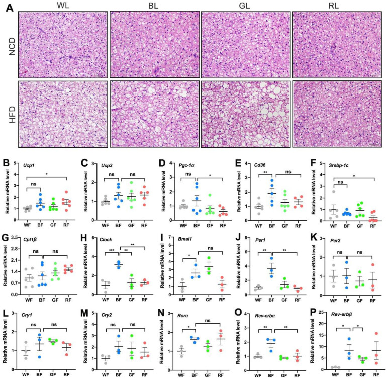
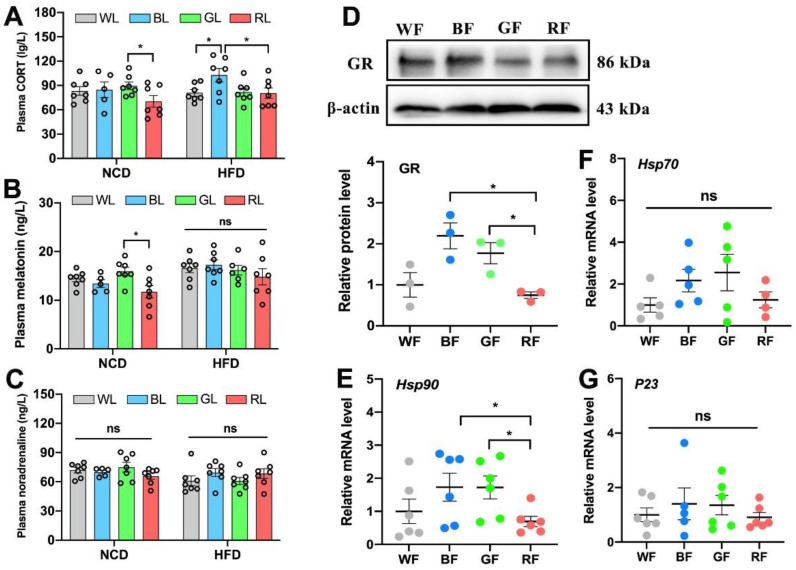
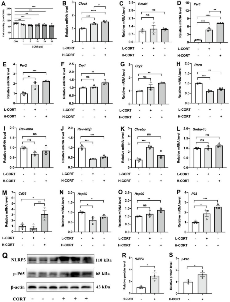
Light pollution worldwide promotes the progression of obesity, which is widely considered a consequence of circadian rhythm disruptions. However, the role of environmental light wavelength in mammalian obesity is not fully understood. Herein, mice fed a normal chow diet (NCD) or a high-fat diet (HFD) were exposed to daytime white (WL), blue (BL), green (GL), and red light (RL) for 8 weeks. Compared with WL and RL, BL significantly increased weight gain and white adipose tissue (WAT) weight, and it disrupted glucose homeostasis in mice fed with HFD but not NCD. The analysis of WAT found that BL significantly aggravated HFD-induced WAT hypertrophy, with a decrease in IL-10 and an increase in NLRP3, p-P65, p-IκB, TLR4, Cd36, Chrebp, Srebp-1c, Fasn, and Cpt1β relative to WL or RL. More interestingly, BL upregulated the expression of circadian clocks in the WAT, including Clock, Bmal1, Per1, Cry1, Cry2, Rorα, Rev-erbα, and Rev-erbβ compared with WL or RL. However, most of the changes had no statistical difference between BL and GL. Mechanistically, BL significantly increased plasma corticosterone (CORT) levels and glucocorticoid receptors in the WAT, which may account for the changes in circadian clocks. Further, in vitro study confirmed that CORT treatment did promote the expression of circadian clocks in 3T3-L1 cells, accompanied by an increase in Chrebp, Cd36, Hsp90, P23, NLRP3, and p-P65. Thus, daily BL, rather than RL exposure-induced CORT elevation, may drive changes in the WAT circadian clocks, ultimately exacerbating lipid dysmetabolism and adipocytic hypertrophy in the HFD-fed mice.
全球光污染促进肥胖的发展,而肥胖被广泛认为是昼夜节律紊乱的结果。然而,环境光波长在哺乳动物肥胖中的作用尚不完全清楚。在此,我们将正常饮食(NCD)或高脂肪饮食(HFD)喂养的小鼠暴露于白天的白光(WL)、蓝光(BL)、绿光(GL)和红光(RL)下 8 周。与 WL 和 RL 相比,BL 显著增加了 HFD 喂养小鼠的体重增加和白色脂肪组织(WAT)重量,并破坏了 HFD 喂养小鼠的葡萄糖稳态,但对 NCD 喂养小鼠没有影响。对 WAT 的分析发现,BL 显著加重了 HFD 诱导的 WAT 肥大,与 WL 或 RL 相比,IL-10 降低,NLRP3、p-P65、p-IκB、TLR4、Cd36、Chrebp、Srebp-1c、Fasn 和 Cpt1β 增加。更有趣的是,与 WL 或 RL 相比,BL 上调了 WAT 中昼夜节律钟的表达,包括 Clock、Bmal1、Per1、Cry1、Cry2、Rorα、Rev-erbα 和 Rev-erbβ。然而,BL 与 GL 之间的大多数变化在统计学上没有差异。从机制上讲,BL 显著增加了 WAT 中的血浆皮质酮(CORT)水平和糖皮质激素受体,这可能是昼夜节律钟变化的原因。此外,体外研究证实 CORT 处理确实促进了 3T3-L1 细胞中昼夜节律钟的表达,伴随着 Chrebp、Cd36、Hsp90、P23、NLRP3 和 p-P65 的增加。因此,与 RL 暴露诱导的 CORT 升高相比,每天的 BL 可能会导致 WAT 昼夜节律钟的变化,最终加剧 HFD 喂养小鼠的脂质代谢紊乱和脂肪细胞肥大。