Stephenson School of Biomedical Engineering, The University of Oklahoma, Norman, OK 73019, United States of America.
Institute for Biomedical Engineering, Science and Technology, The University of Oklahoma, Norman, OK 73019, United States of America.
J Neural Eng. 2023 Jan 18;20(1):016012. doi: 10.1088/1741-2552/acaccb.
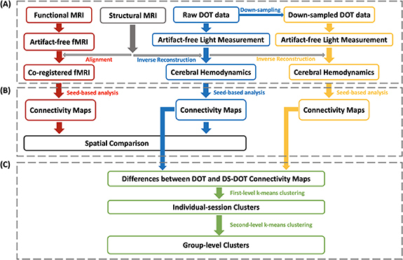
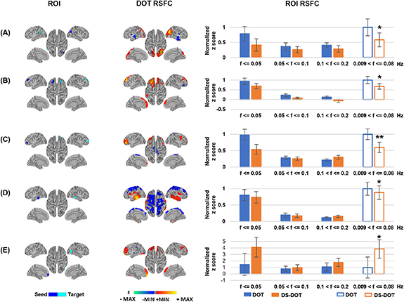
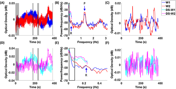
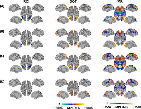
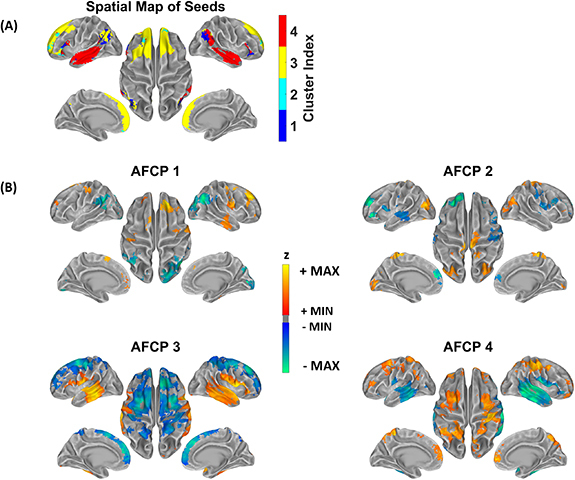
. Spontaneous fluctuations of cerebral hemodynamics measured by functional magnetic resonance imaging (fMRI) are widely used to study the network organization of the brain. The temporal correlations among the ultra-slow, <0.1 Hz fluctuations across the brain regions are interpreted as functional connectivity maps and used for diagnostics of neurological disorders. However, despite the interest narrowed in the ultra-slow fluctuations, hemodynamic activity that exists beyond the ultra-slow frequency range could contribute to the functional connectivity, which remains unclear.. In the present study, we have measured the brain-wide hemodynamics in the human participants with functional near-infrared spectroscopy (fNIRS) in a whole-head, cap-based and high-density montage at a sampling rate of 6.25 Hz. In addition, we have acquired resting state fMRI scans in the same group of participants for cross-modal evaluation of the connectivity maps. Then fNIRS data were deliberately down-sampled to a typical fMRI sampling rate of ∼0.5 Hz and the resulted differential connectivity maps were subject to a k-means clustering.. Our diffuse optical topographical analysis of fNIRS data have revealed a default mode network (DMN) in the spontaneous deoxygenated and oxygenated hemoglobin changes, which remarkably resemble the same fMRI network derived from participants. Moreover, we have shown that the aliased activities in the down-sampled optical signals have altered the connectivity patterns, resulting in a network organization of aliased functional connectivity in the cerebral hemodynamics.The results have for the first time demonstrated that fNIRS as a broadly accessible modality can image the resting-state functional connectivity in the posterior midline, prefrontal and parietal structures of the DMN in the human brain, in a consistent pattern with fMRI. Further empowered by the fast sampling rate of fNIRS, our findings suggest the presence of aliased connectivity in the current understanding of the human brain organization.
. 功能磁共振成像(fMRI)测量的脑血流动力学自发性波动被广泛用于研究大脑的网络组织。大脑区域之间超慢(<0.1 Hz)波动的时间相关性被解释为功能连接图,并用于神经障碍的诊断。然而,尽管人们对超慢波动的兴趣越来越浓厚,但超出超慢频率范围的血流动力学活动可能会对功能连接产生影响,这一点尚不清楚。. 在本研究中,我们使用功能近红外光谱(fNIRS)以全头、帽式和高密度装置在 6.25 Hz 的采样率测量了人类参与者的全脑血流动力学。此外,我们在同一组参与者中获取了静息状态 fMRI 扫描,以进行连接图的跨模态评估。然后,我们将 fNIRS 数据故意下采样到典型的 fMRI 采样率约为 0.5 Hz,并对得到的差分连接图进行 k-均值聚类分析。. 我们对 fNIRS 数据的漫射光学拓扑分析揭示了自发去氧和氧合血红蛋白变化中的默认模式网络(DMN),这与从参与者中得出的相同 fMRI 网络非常相似。此外,我们表明,下采样光信号中的伪影活动改变了连接模式,导致大脑血流动力学中伪影功能连接的网络组织。研究结果首次表明,fNIRS 作为一种广泛可及的模态,可以在与 fMRI 一致的模式下,对人类大脑中 DMN 的后中线、前额叶和顶叶结构的静息状态功能连接进行成像。fNIRS 的快速采样率进一步增强了我们的发现,表明在当前对人类大脑组织的理解中存在伪影连接。