• 文献检索
  • 文档翻译
  • 深度研究
  • 学术资讯
  • Suppr Zotero 插件Zotero 插件
  • 邀请有礼
  • 套餐&价格
  • 历史记录
应用&插件
Suppr Zotero 插件Zotero 插件浏览器插件Mac 客户端Windows 客户端微信小程序
定价
高级版会员购买积分包购买API积分包
服务
文献检索文档翻译深度研究API 文档MCP 服务
关于我们
关于 Suppr公司介绍联系我们用户协议隐私条款
关注我们

Suppr 超能文献

核心技术专利:CN118964589B侵权必究
粤ICP备2023148730 号-1Suppr @ 2026

文献检索

告别复杂PubMed语法,用中文像聊天一样搜索,搜遍4000万医学文献。AI智能推荐,让科研检索更轻松。

立即免费搜索

文件翻译

保留排版,准确专业,支持PDF/Word/PPT等文件格式,支持 12+语言互译。

免费翻译文档

深度研究

AI帮你快速写综述,25分钟生成高质量综述,智能提取关键信息,辅助科研写作。

立即免费体验

髋关节骨关节炎:X 线摄影仍有必要吗?

Osteoarthritis of the hip: is radiography still needed?

机构信息

Department of Diagnostic and Interventional Radiology, Hôpital Libanais Geitaoui CHU, Beyrouth, 1100, Achrafieh, Lebanon.

Department of Radiology, Cliniques CHC Montlégia, Boulevard Patience Et Beaujonc 2, 4000, Liège, Belgium.

出版信息

Skeletal Radiol. 2023 Nov;52(11):2259-2270. doi: 10.1007/s00256-022-04270-8. Epub 2022 Dec 20.

DOI:10.1007/s00256-022-04270-8
PMID:36538067
原文链接:https://pmc.ncbi.nlm.nih.gov/articles/PMC10509135/
Abstract

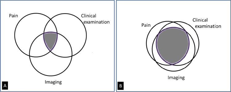
Diagnosis of hip osteoarthritis (OA) is based on clinical arguments, and medical imaging is obtained to confirm the diagnosis and rule out other possible sources of pain. Conventional radiographs are recommended as the first line imaging modality to investigate chronic hip pain. They should be obtained in a rigorous technique that includes an antero-posterior (AP) radiograph of the pelvis. The choice of the appropriate lateral view depends on the clinical indication, Lequesne's false profile being valuable in the assessment of OA. Magnetic resonance imaging (MRI) is more sensitive to detect joint effusion/synovitis, cartilage, labral, and bone marrow lesions. However, structural joint changes are frequent in asymptomatic population and neither radiographs nor MRI have shown a good correlation with pain and functional impairment. MRI seems to be more suitable than radiographs as a biomarker for clinical trials addressing early OA. The absence of a validated MR biomarker of early OA, together with issues related to machine availability and MRI protocol repeatability, prevent the widespread use of MRI in clinical trials.

摘要

髋关节骨关节炎(OA)的诊断基于临床依据,并且获取医学影像学检查来确认诊断并排除其他可能的疼痛来源。常规 X 线片被推荐作为研究慢性髋关节疼痛的一线影像学方法。它们应该在严格的技术下获得,包括骨盆的前后位(AP)X 线片。适当的侧位片的选择取决于临床指征,Lequesne 的假性侧面位在 OA 的评估中具有价值。磁共振成像(MRI)对检测关节积液/滑膜炎、软骨、盂唇和骨髓病变更敏感。然而,结构关节变化在无症状人群中很常见,并且 X 线片和 MRI 都没有显示出与疼痛和功能障碍的良好相关性。MRI 似乎比 X 线片更适合作为临床试验中针对早期 OA 的生物标志物。缺乏早期 OA 的经验证的 MRI 生物标志物,以及与机器可用性和 MRI 协议可重复性相关的问题,阻碍了 MRI 在临床试验中的广泛应用。

https://cdn.ncbi.nlm.nih.gov/pmc/blobs/6512/10509135/c7f1351ab2b8/256_2022_4270_Fig7_HTML.jpg
https://cdn.ncbi.nlm.nih.gov/pmc/blobs/6512/10509135/25ba3968a2eb/256_2022_4270_Fig1_HTML.jpg
https://cdn.ncbi.nlm.nih.gov/pmc/blobs/6512/10509135/ad511a98c136/256_2022_4270_Fig2_HTML.jpg
https://cdn.ncbi.nlm.nih.gov/pmc/blobs/6512/10509135/4f1fb6c1d94e/256_2022_4270_Fig3_HTML.jpg
https://cdn.ncbi.nlm.nih.gov/pmc/blobs/6512/10509135/6c24fe7034ee/256_2022_4270_Fig4_HTML.jpg
https://cdn.ncbi.nlm.nih.gov/pmc/blobs/6512/10509135/fe5499f24420/256_2022_4270_Fig5_HTML.jpg
https://cdn.ncbi.nlm.nih.gov/pmc/blobs/6512/10509135/74b159e636d3/256_2022_4270_Fig6_HTML.jpg
https://cdn.ncbi.nlm.nih.gov/pmc/blobs/6512/10509135/c7f1351ab2b8/256_2022_4270_Fig7_HTML.jpg

相似文献

1
Osteoarthritis of the hip: is radiography still needed?髋关节骨关节炎:X 线摄影仍有必要吗?
Skeletal Radiol. 2023 Nov;52(11):2259-2270. doi: 10.1007/s00256-022-04270-8. Epub 2022 Dec 20.
2
[When should MRI for knee or hip osteoarthritis should be performed?].[膝关节或髋关节骨关节炎何时应进行磁共振成像(MRI)检查?]
Rev Prat. 2012 May;62(5):625-9.
3
Recommendations for the use of new methods to assess the efficacy of disease-modifying drugs in the treatment of osteoarthritis.关于使用新方法评估改善病情药物治疗骨关节炎疗效的建议。
Osteoarthritis Cartilage. 2004 Apr;12(4):263-8. doi: 10.1016/j.joca.2004.01.006.
4
Association between hip and knee cartilage measured using radiographs and magnetic resonance imaging: the Tasmanian Older Adult Cohort Study.基于放射影像和磁共振成像的髋关节和膝关节软骨关联性研究:塔斯马尼亚中老年队列研究。
Rheumatology (Oxford). 2013 Nov;52(11):2009-15. doi: 10.1093/rheumatology/ket243. Epub 2013 Jul 30.
5
Imaging of Osteoarthritis of the Hip.髋关节骨关节炎的影像学表现。
Radiol Clin North Am. 2022 Jul;60(4):617-628. doi: 10.1016/j.rcl.2022.03.005. Epub 2022 May 21.
6
Semiquantitative Imaging Biomarkers of Knee Osteoarthritis Progression: Data From the Foundation for the National Institutes of Health Osteoarthritis Biomarkers Consortium.膝关节骨关节炎进展的半定量成像生物标志物:来自美国国立卫生研究院骨关节炎生物标志物联盟的资料。
Arthritis Rheumatol. 2016 Oct;68(10):2422-31. doi: 10.1002/art.39731.
7
Evaluation of the 'Lequesne's false profile' of the hip in patients with hip osteoarthritis.髋骨关节炎患者髋关节“Lequesne假轮廓”的评估。
Osteoarthritis Cartilage. 1999 May;7(3):295-300. doi: 10.1053/joca.1998.0203.
8
Associations between suprapatellar pouch effusion-synovitis, serum cartilage oligomeric matrix protein, high sensitivity C-reaction protein, knee symptom, and joint structural changes in patients with knee osteoarthritis.髌上囊积液-滑膜炎、血清软骨寡聚基质蛋白、高敏 C 反应蛋白与膝关节骨关节炎患者膝关节症状及关节结构变化的相关性。
Clin Rheumatol. 2020 May;39(5):1663-1670. doi: 10.1007/s10067-019-04905-7. Epub 2020 Jan 3.
9
Predictive value of MRI features for development of radiographic osteoarthritis in a cohort of participants with pre-radiographic knee osteoarthritis-the CHECK study.MRI特征对膝关节X线前期骨关节炎队列中X线骨关节炎发生的预测价值——CHECK研究
Rheumatology (Oxford). 2017 Jan;56(1):113-120. doi: 10.1093/rheumatology/kew368. Epub 2016 Oct 25.
10
Clinical and radiological evaluation of hip, knee and hand osteoarthritis.髋、膝和手部骨关节炎的临床与影像学评估。
Aging Clin Exp Res. 2003 Oct;15(5):380-90. doi: 10.1007/BF03327359.

引用本文的文献

1
Radiographic evaluation of the painful adolescent and young adult hip.疼痛性青少年及青年成人髋关节的影像学评估
J Pediatr Soc North Am. 2024 Apr 3;7:100039. doi: 10.1016/j.jposna.2024.100039. eCollection 2024 May.
2
Comparison of a New Radiographic Technique with MRI Measurements for Tibial Tunnel Evaluation in ACL Reconstruction.一种用于前交叉韧带重建中胫骨隧道评估的新影像学技术与磁共振成像测量的比较
Diagnostics (Basel). 2025 May 14;15(10):1237. doi: 10.3390/diagnostics15101237.
3
A quantitative analysis of symmetry on standard anteroposterior pelvic X-ray.

本文引用的文献

1
Proportion of patients with hip osteoarthritis in primary care identified by differing clinical criteria: a cross-sectional study of 4699 patients.根据不同临床标准确定的基层医疗中髋骨关节炎患者的比例:一项对4699名患者的横断面研究
Osteoarthr Cartil Open. 2020 Oct 13;2(4):100111. doi: 10.1016/j.ocarto.2020.100111. eCollection 2020 Dec.
2
Global, regional, and national burdens of hip osteoarthritis from 1990 to 2019: estimates from the 2019 Global Burden of Disease Study.全球、区域和国家髋骨骨关节炎负担:来自 2019 年全球疾病负担研究的估计。
Arthritis Res Ther. 2022 Jan 3;24(1):8. doi: 10.1186/s13075-021-02705-6.
3
标准骨盆前后位X线片对称性的定量分析。
Acta Orthop Traumatol Turc. 2025 Apr 29;59(2):122-128. doi: 10.5152/j.aott.2025.24033.
4
Predictive and classification capabilities of the timed up and go as a physical performance measure in hip osteoarthritis: a retrospective study of 606 patients.《 timed up and go 在髋骨关节炎中的物理表现测量中的预测和分类能力:606 名患者的回顾性研究》。
Arch Orthop Trauma Surg. 2024 Sep;144(9):4171-4178. doi: 10.1007/s00402-024-05505-0. Epub 2024 Sep 6.
5
Holistic vibrational spectromics assessment of human cartilage for osteoarthritis diagnosis.用于骨关节炎诊断的人体软骨的整体振动光谱评估。
Biomed Opt Express. 2024 Jun 13;15(7):4264-4280. doi: 10.1364/BOE.520171. eCollection 2024 Jul 1.
6
The Invasive Management of Pain: Diagnosis and New Treatment Options.疼痛的侵入性管理:诊断与新的治疗选择
Cureus. 2023 Jul 31;15(7):e42717. doi: 10.7759/cureus.42717. eCollection 2023 Jul.
Ossification of the acetabular rim: a highly prevalent finding in asymptomatic non-osteoarthritic hips of all ages.
髋臼缘骨化:各年龄段无症状非骨关节炎髋关节中非常普遍的发现。
Eur Radiol. 2021 Sep;31(9):6802-6809. doi: 10.1007/s00330-021-07750-y. Epub 2021 Mar 13.
4
Acetabular Cartilage Thickness Differs Among Cam, Pincer, or Mixed-Type Femoroacetabular Impingement: A Descriptive Study Using Ultrasonic Measurements During Surgical Hip Dislocation.髋臼软骨厚度在凸轮型、钳夹型或混合型股骨髋臼撞击症中存在差异:一项使用超声测量技术在髋关节脱位手术中进行的描述性研究。
Cartilage. 2021 Dec;13(2_suppl):465S-475S. doi: 10.1177/1947603521990879. Epub 2021 Feb 8.
5
The Lisbon Agreement on Femoroacetabular Impingement Imaging-part 1: overview.《髋关节撞击综合征影像学检查的里斯本协议》-第 1 部分:概述。
Eur Radiol. 2020 Oct;30(10):5281-5297. doi: 10.1007/s00330-020-06822-9. Epub 2020 Jul 17.
6
The Italian Society for Rheumatology clinical practice guidelines for the diagnosis and management of knee, hip and hand osteoarthritis.意大利风湿病学会关于膝关节、髋关节和手部骨关节炎诊断与管理的临床实践指南。
Reumatismo. 2019 Sep 23;71(S1):5-21. doi: 10.4081/reumatismo.2019.1188.
7
Does This Patient Have Hip Osteoarthritis?: The Rational Clinical Examination Systematic Review.这位患者是否患有髋骨骨关节炎?:合理临床检查系统评价。
JAMA. 2019 Dec 17;322(23):2323-2333. doi: 10.1001/jama.2019.19413.
8
Conventional Radiography of the Hip Revisited: Correlation with Advanced Imaging.髋关节传统放射成像再探讨:与先进成像技术的相关性
Magn Reson Imaging Clin N Am. 2019 Nov;27(4):661-683. doi: 10.1016/j.mric.2019.07.002.
9
Position paper on magnetic resonance imaging protocols in the musculoskeletal system (excluding the spine) by the Italian College of Musculoskeletal Radiology.意大利肌肉骨骼放射学会关于肌肉骨骼系统(不包括脊柱)磁共振成像协议的立场文件。
Radiol Med. 2019 Jun;124(6):522-538. doi: 10.1007/s11547-019-00992-3. Epub 2019 Jan 28.
10
Classifications in Brief: Tönnis Classification of Hip Osteoarthritis.简要分类:髋关节骨关节炎的托尼分类法。
Clin Orthop Relat Res. 2018 Aug;476(8):1680-1684. doi: 10.1097/01.blo.0000534679.75870.5f.