• 文献检索
  • 文档翻译
  • 深度研究
  • 学术资讯
  • Suppr Zotero 插件Zotero 插件
  • 邀请有礼
  • 套餐&价格
  • 历史记录
应用&插件
Suppr Zotero 插件Zotero 插件浏览器插件Mac 客户端Windows 客户端微信小程序
定价
高级版会员购买积分包购买API积分包
服务
文献检索文档翻译深度研究API 文档MCP 服务
关于我们
关于 Suppr公司介绍联系我们用户协议隐私条款
关注我们

Suppr 超能文献

核心技术专利:CN118964589B侵权必究
粤ICP备2023148730 号-1Suppr @ 2026

文献检索

告别复杂PubMed语法,用中文像聊天一样搜索,搜遍4000万医学文献。AI智能推荐,让科研检索更轻松。

立即免费搜索

文件翻译

保留排版,准确专业,支持PDF/Word/PPT等文件格式,支持 12+语言互译。

免费翻译文档

深度研究

AI帮你快速写综述,25分钟生成高质量综述,智能提取关键信息,辅助科研写作。

立即免费体验

一种基于新型生成对抗网络的自动脑肿瘤分割方法。

A Novel Generative Adversarial Network-Based Approach for Automated Brain Tumour Segmentation.

机构信息

School of Computer Science, University of Petroleum and Energy Studies (UPES), Dehradun 248007, India.

Persistent Systems, India 411057, India.

出版信息

Medicina (Kaunas). 2023 Jan 6;59(1):119. doi: 10.3390/medicina59010119.

DOI:10.3390/medicina59010119
PMID:36676743
原文链接:https://pmc.ncbi.nlm.nih.gov/articles/PMC9863906/
Abstract

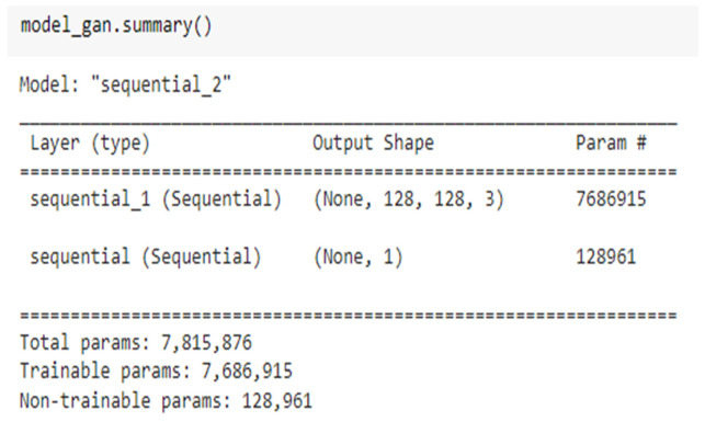
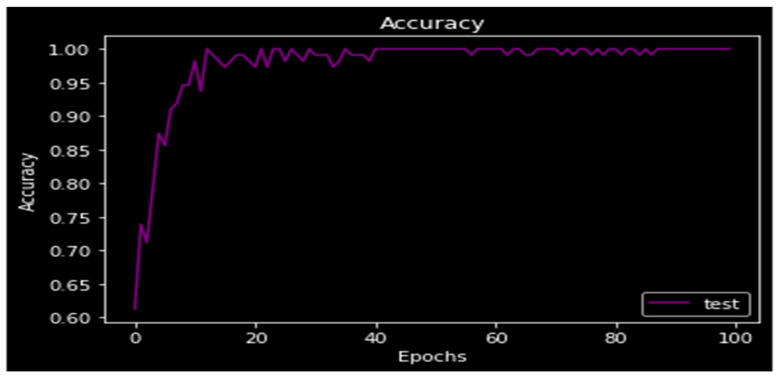
: Medical image segmentation is more complicated and demanding than ordinary image segmentation due to the density of medical pictures. A brain tumour is the most common cause of high mortality. : Extraction of tumorous cells is particularly difficult due to the differences between tumorous and non-tumorous cells. In ordinary convolutional neural networks, local background information is restricted. As a result, previous deep learning algorithms in medical imaging have struggled to detect anomalies in diverse cells. : As a solution to this challenge, a deep convolutional generative adversarial network for tumour segmentation from brain Magnetic resonance Imaging (MRI) images is proposed. A generator and a discriminator are the two networks that make up the proposed model. This network focuses on tumour localisation, noise-related issues, and social class disparities. : Dice Score Coefficient (DSC), Peak Signal to Noise Ratio (PSNR), and Structural Index Similarity (SSIM) are all generally 0.894, 62.084 dB, and 0.88912, respectively. The model's accuracy has improved to 97 percent, and its loss has reduced to 0.012. : Experiments reveal that the proposed approach may successfully segment tumorous and benign tissues. As a result, a novel brain tumour segmentation approach has been created.

摘要

医学图像分割比普通图像分割复杂且要求更高,因为医学图像密度较高。脑肿瘤是导致高死亡率的最常见原因。由于肿瘤细胞和非肿瘤细胞之间存在差异,因此提取肿瘤细胞尤其困难。在普通卷积神经网络中,局部背景信息受到限制。因此,以前的医学影像深度学习算法难以检测不同细胞中的异常。为了解决这个挑战,提出了一种用于从脑磁共振成像(MRI)图像中分割肿瘤的深度卷积生成对抗网络。所提出的模型由生成器和鉴别器两个网络组成。该网络专注于肿瘤定位、与噪声相关的问题和社会阶层差异。Dice 得分系数(DSC)、峰值信噪比(PSNR)和结构相似性指数(SSIM)分别为 0.894、62.084 dB 和 0.88912。模型的准确率提高到 97%,损失降低到 0.012。实验表明,所提出的方法可以成功分割肿瘤和良性组织。因此,创建了一种新的脑肿瘤分割方法。

https://cdn.ncbi.nlm.nih.gov/pmc/blobs/d67c/9863906/7d35be75f61d/medicina-59-00119-g011.jpg
https://cdn.ncbi.nlm.nih.gov/pmc/blobs/d67c/9863906/e1288102d8bf/medicina-59-00119-g001.jpg
https://cdn.ncbi.nlm.nih.gov/pmc/blobs/d67c/9863906/0059c55adb78/medicina-59-00119-g002.jpg
https://cdn.ncbi.nlm.nih.gov/pmc/blobs/d67c/9863906/13286b07bc66/medicina-59-00119-g003.jpg
https://cdn.ncbi.nlm.nih.gov/pmc/blobs/d67c/9863906/c2b9f8699e01/medicina-59-00119-g004.jpg
https://cdn.ncbi.nlm.nih.gov/pmc/blobs/d67c/9863906/eba4f38eb1d6/medicina-59-00119-g005.jpg
https://cdn.ncbi.nlm.nih.gov/pmc/blobs/d67c/9863906/37a17afa4e23/medicina-59-00119-g006.jpg
https://cdn.ncbi.nlm.nih.gov/pmc/blobs/d67c/9863906/0c8f8890c0f9/medicina-59-00119-g007.jpg
https://cdn.ncbi.nlm.nih.gov/pmc/blobs/d67c/9863906/d023e742a018/medicina-59-00119-g008.jpg
https://cdn.ncbi.nlm.nih.gov/pmc/blobs/d67c/9863906/4e08c3dd45ee/medicina-59-00119-g009.jpg
https://cdn.ncbi.nlm.nih.gov/pmc/blobs/d67c/9863906/20d0a34514ae/medicina-59-00119-g010.jpg
https://cdn.ncbi.nlm.nih.gov/pmc/blobs/d67c/9863906/7d35be75f61d/medicina-59-00119-g011.jpg

相似文献

1
A Novel Generative Adversarial Network-Based Approach for Automated Brain Tumour Segmentation.一种基于新型生成对抗网络的自动脑肿瘤分割方法。
Medicina (Kaunas). 2023 Jan 6;59(1):119. doi: 10.3390/medicina59010119.
2
Brain tumor classification for MRI images using dual-discriminator conditional generative adversarial network.基于双鉴别器条件生成对抗网络的 MRI 图像脑肿瘤分类。
Electromagn Biol Med. 2024 Apr 2;43(1-2):81-94. doi: 10.1080/15368378.2024.2321352. Epub 2024 Mar 10.
3
Multi-Modal Brain Tumor Data Completion Based on Reconstruction Consistency Loss.基于重建一致性损失的多模态脑肿瘤数据补全。
J Digit Imaging. 2023 Aug;36(4):1794-1807. doi: 10.1007/s10278-022-00697-6. Epub 2023 Mar 1.
4
Pseudo-CT generation from multi-parametric MRI using a novel multi-channel multi-path conditional generative adversarial network for nasopharyngeal carcinoma patients.基于新型多通道多路径条件生成对抗网络的多参数 MRI 伪 CT 生成用于鼻咽癌患者。
Med Phys. 2020 Apr;47(4):1750-1762. doi: 10.1002/mp.14062. Epub 2020 Feb 21.
5
Shape constrained fully convolutional DenseNet with adversarial training for multiorgan segmentation on head and neck CT and low-field MR images.基于对抗训练的形状约束全卷积 DenseNet 用于头颈部 CT 和低场 MR 图像多器官分割。
Med Phys. 2019 Jun;46(6):2669-2682. doi: 10.1002/mp.13553. Epub 2019 May 6.
6
Automated cartilage and meniscus segmentation of knee MRI with conditional generative adversarial networks.基于条件生成对抗网络的膝关节磁共振成像中软骨和半月板的自动分割
Magn Reson Med. 2020 Jul;84(1):437-449. doi: 10.1002/mrm.28111. Epub 2019 Dec 2.
7
Improved brain metastases segmentation using generative adversarial network and conditional random field optimization mask R-CNN.利用生成对抗网络和条件随机场优化掩模 R-CNN 提高脑转移瘤分割效果。
Med Phys. 2024 Sep;51(9):5990-6001. doi: 10.1002/mp.17176. Epub 2024 May 22.
8
Medical image synthesis via conditional GANs: Application to segmenting brain tumours.基于条件生成对抗网络的医学图像合成:在脑肿瘤分割中的应用。
Comput Biol Med. 2024 Mar;170:107982. doi: 10.1016/j.compbiomed.2024.107982. Epub 2024 Jan 18.
9
DualMMP-GAN: Dual-scale multi-modality perceptual generative adversarial network for medical image segmentation.双尺度多模态感知生成对抗网络用于医学图像分割。
Comput Biol Med. 2022 May;144:105387. doi: 10.1016/j.compbiomed.2022.105387. Epub 2022 Mar 12.
10
Self-attention-based generative adversarial network optimized with color harmony algorithm for brain tumor classification.基于自注意力的生成对抗网络,结合颜色调和算法,用于脑肿瘤分类。
Electromagn Biol Med. 2024 Apr 2;43(1-2):31-45. doi: 10.1080/15368378.2024.2312363. Epub 2024 Feb 18.

引用本文的文献

1
MSA-Net: a multi-scale and adversarial learning network for segmenting bone metastases in low-resolution SPECT imaging.MSA-Net:一种用于在低分辨率单光子发射计算机断层扫描成像中分割骨转移灶的多尺度对抗学习网络。
EJNMMI Phys. 2025 Jul 24;12(1):72. doi: 10.1186/s40658-025-00785-w.
2
Improving the Generalizability of Deep Learning for T2-Lesion Segmentation of Gliomas in the Post-Treatment Setting.提高深度学习在治疗后胶质瘤T2病变分割中的通用性。
Bioengineering (Basel). 2024 May 16;11(5):497. doi: 10.3390/bioengineering11050497.

本文引用的文献

1
Brain tumor image generation using an aggregation of GAN models with style transfer.基于具有风格迁移的 GAN 模型聚合的脑肿瘤图像生成。
Sci Rep. 2022 Jun 1;12(1):9141. doi: 10.1038/s41598-022-12646-y.
2
Correcting and reweighting false label masks in brain tumor segmentation.纠正和重新加权脑肿瘤分割中的错误标签掩模。
Med Phys. 2021 Jan;48(1):169-177. doi: 10.1002/mp.14480. Epub 2020 Nov 29.
3
CTumorGAN: a unified framework for automatic computed tomography tumor segmentation.CTumorGAN:用于自动计算机断层扫描肿瘤分割的统一框架。
Eur J Nucl Med Mol Imaging. 2020 Sep;47(10):2248-2268. doi: 10.1007/s00259-020-04781-3. Epub 2020 Mar 28.
4
Tumor-aware, Adversarial Domain Adaptation from CT to MRI for Lung Cancer Segmentation.用于肺癌分割的从CT到MRI的肿瘤感知对抗域适应
Med Image Comput Comput Assist Interv. 2018 Sep;11071:777-785. doi: 10.1007/978-3-030-00934-2_86.
5
Advancing The Cancer Genome Atlas glioma MRI collections with expert segmentation labels and radiomic features.利用专家分割标签和放射组学特征推进癌症基因组图谱胶质细胞瘤 MRI 数据集。
Sci Data. 2017 Sep 5;4:170117. doi: 10.1038/sdata.2017.117.
6
Image Analysis for MRI Based Brain Tumor Detection and Feature Extraction Using Biologically Inspired BWT and SVM.基于生物启发的BWT和SVM的MRI脑肿瘤检测与特征提取的图像分析
Int J Biomed Imaging. 2017;2017:9749108. doi: 10.1155/2017/9749108. Epub 2017 Mar 6.
7
Brain tumor segmentation with Deep Neural Networks.基于深度神经网络的脑肿瘤分割。
Med Image Anal. 2017 Jan;35:18-31. doi: 10.1016/j.media.2016.05.004. Epub 2016 May 19.
8
The Multimodal Brain Tumor Image Segmentation Benchmark (BRATS).多模态脑肿瘤图像分割基准(BRATS)。
IEEE Trans Med Imaging. 2015 Oct;34(10):1993-2024. doi: 10.1109/TMI.2014.2377694. Epub 2014 Dec 4.
9
New variants of a method of MRI scale standardization.磁共振成像(MRI)尺度标准化方法的新变体
IEEE Trans Med Imaging. 2000 Feb;19(2):143-50. doi: 10.1109/42.836373.