Chadchan Sangappa B, Naik Sumanta K, Popli Pooja, Talwar Chandni, Putluri Satwikreddy, Ambati Chandrasekhar R, Lint Michael A, Kau Andrew L, Stallings Christina L, Kommagani Ramakrishna
Department of Pathology and Immunology, Baylor College of Medicine, One Baylor Plaza, Houston, TX, 77030, USA.
Department of Molecular Microbiology, Washington University School of Medicine, St. Louis, MO, 63110, USA.
Cell Death Discov. 2023 Jan 25;9(1):28. doi: 10.1038/s41420-023-01309-0.
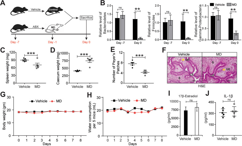
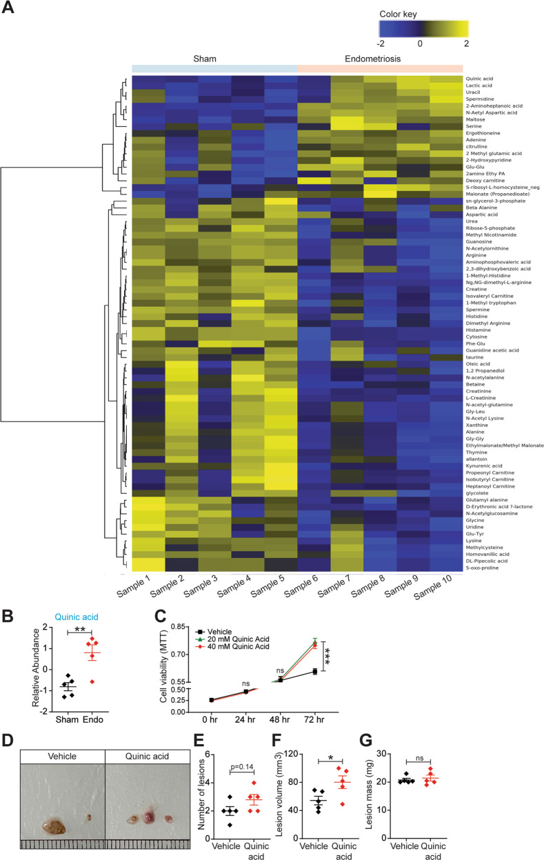
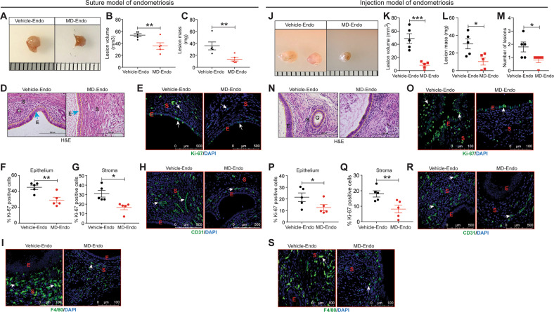
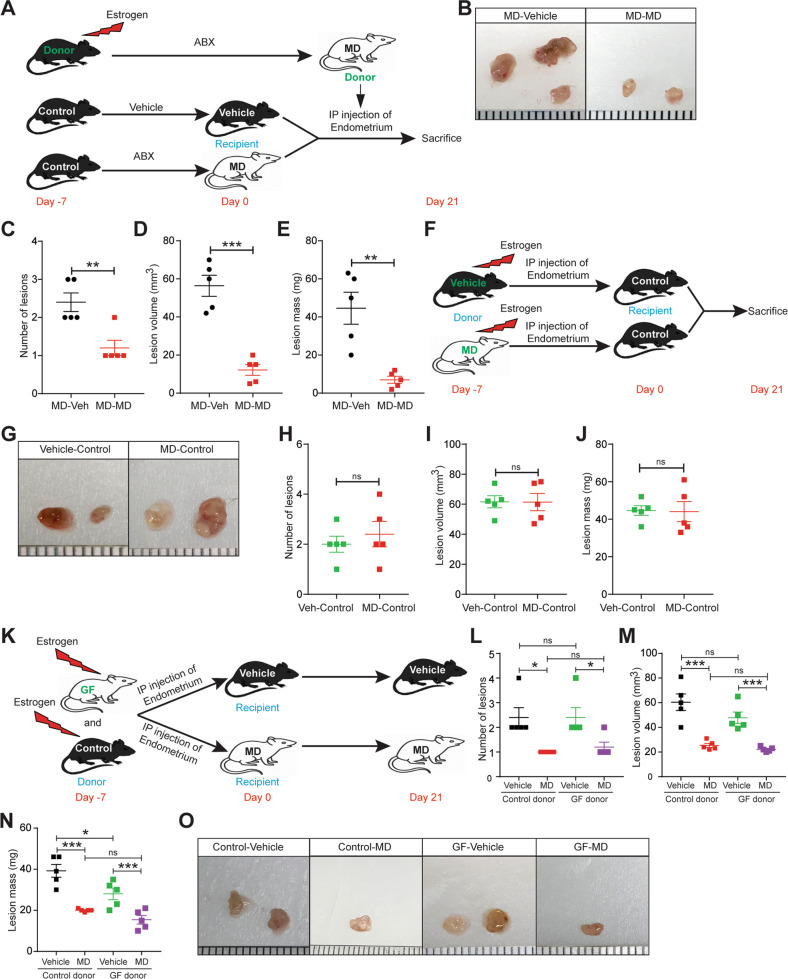
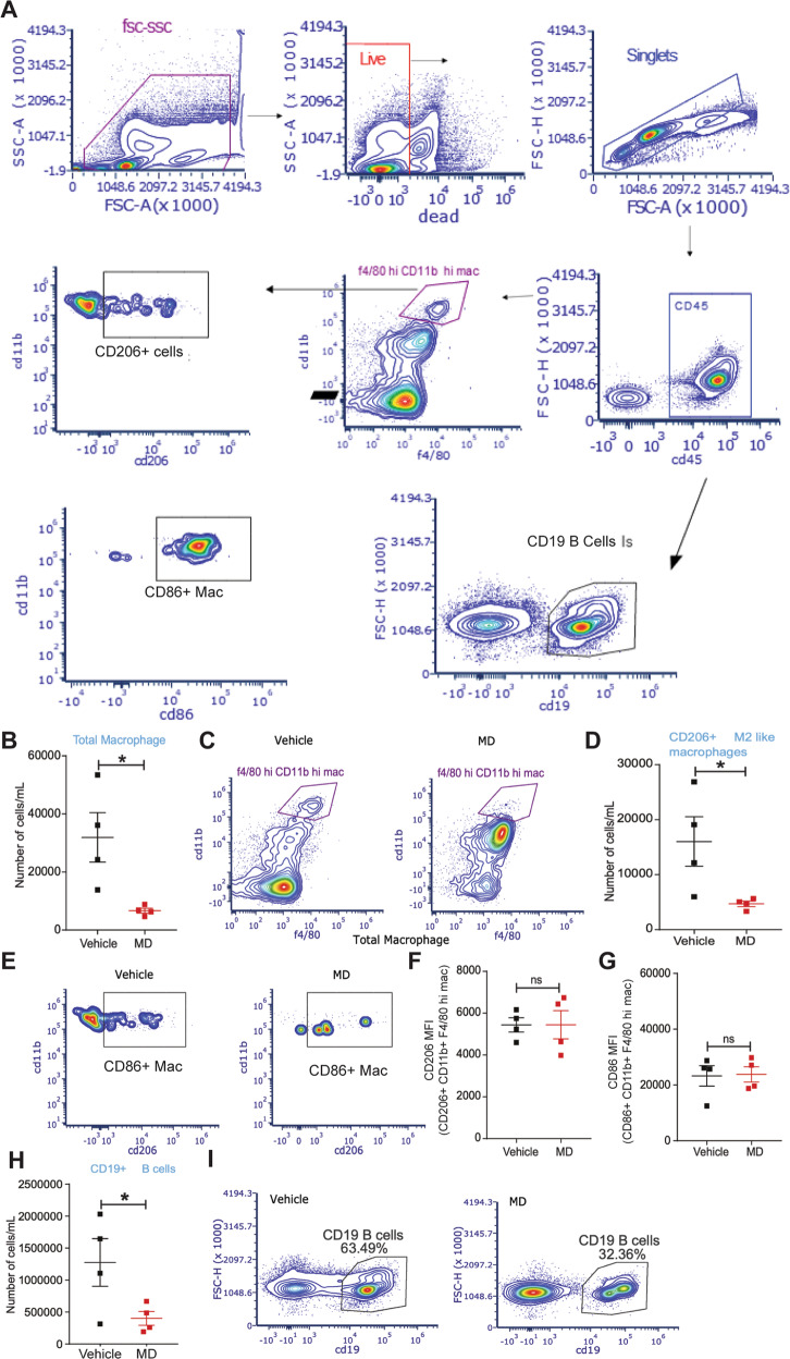
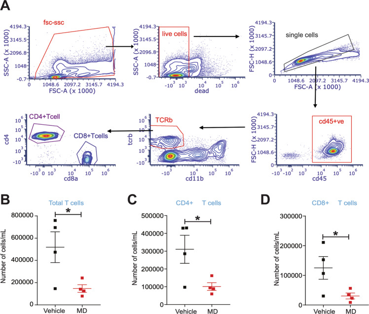
Endometriosis is a pathological condition of the female reproductive tract characterized by the existence of endometrium-like tissue at ectopic sites, affecting 10% of women between the age 15 and 49 in the USA. However, currently there is no reliable non-invasive method to detect the presence of endometriosis without surgery and many women find hormonal therapy and surgery as ineffective in avoiding the recurrences. There is a lack of knowledge on the etiology and the factors that contribute to the development of endometriosis. A growing body of recent evidence suggests an association between gut microbiota and endometriosis pathophysiology. However, the direct impact of microbiota and microbiota-derived metabolites on the endometriosis disease progression is largely unknown. To understand the causal role of gut microbiota and endometriosis, we have implemented a novel model using antibiotic-induced microbiota-depleted (MD) mice to investigate the endometriosis disease progression. Interestingly, we found that MD mice showed reduced endometriotic lesion growth and, the transplantation of gut microbiota by oral gavage of feces from mice with endometriosis rescued the endometriotic lesion growth. Additionally, using germ-free donor mice, we indicated that the uterine microbiota is dispensable for endometriotic lesion growth in mice. Furthermore, we showed that gut microbiota modulates immune cell populations in the peritoneum of lesions-bearing mice. Finally, we found a novel signature of microbiota-derived metabolites that were significantly altered in feces of mice with endometriosis. Finally, we found one the altered metabolite, quinic acid promoted the survival of endometriotic epithelial cells in vitro and lesion growth in vivo, suggesting the disease-promoting potential of microbiota-derived metabolites. In summary, these data suggest that gut microbiota and microbiota-derived metabolome contribute to lesion growth in mice, possibly through immune cell adaptations. Of translational significance, these findings will aid in designing non-invasive diagnostics using stool metabolites for endometriosis.
子宫内膜异位症是一种女性生殖道的病理状况,其特征是在异位部位存在类似子宫内膜的组织,在美国15至49岁的女性中,有10%受其影响。然而,目前尚无可靠的非侵入性方法可在不进行手术的情况下检测子宫内膜异位症的存在,而且许多女性发现激素疗法和手术在避免复发方面效果不佳。目前对子宫内膜异位症的病因及促成其发展的因素尚缺乏了解。最近越来越多的证据表明肠道微生物群与子宫内膜异位症的病理生理之间存在关联。然而,微生物群及其衍生代谢产物对子宫内膜异位症疾病进展的直接影响在很大程度上尚不清楚。为了解肠道微生物群与子宫内膜异位症之间的因果关系,我们采用了一种新型模型,即使用抗生素诱导的微生物群耗竭(MD)小鼠来研究子宫内膜异位症的疾病进展。有趣的是,我们发现MD小鼠的子宫内膜异位病变生长减缓,而通过口服来自患有子宫内膜异位症小鼠的粪便进行肠道微生物群移植可挽救子宫内膜异位病变的生长。此外,利用无菌供体小鼠,我们表明子宫微生物群对小鼠子宫内膜异位病变的生长并非必需。此外,我们还表明肠道微生物群可调节患有病变小鼠腹膜中的免疫细胞群体。最后,我们发现了一种微生物群衍生代谢产物的新特征,这些代谢产物在患有子宫内膜异位症小鼠的粪便中发生了显著变化。最后,我们发现其中一种改变的代谢产物奎尼酸在体外促进了子宫内膜异位上皮细胞的存活,并在体内促进了病变生长,这表明微生物群衍生代谢产物具有促进疾病的潜力。总之,这些数据表明肠道微生物群及其衍生的代谢组可能通过免疫细胞适应促进小鼠病变生长。具有转化意义的是,这些发现将有助于设计利用粪便代谢产物对子宫内膜异位症进行非侵入性诊断的方法。