Department of Radiology, Jiangxi Provincial People's Hospital, The First Affiliated Hospital of Nanchang Medical College, 152 Aiguo Road, Nanchang, 330006, China.
Department of Gynecology, Jiangxi Provincial People's Hospital, The First Affiliated Hospital of Nanchang Medical College, Nanchang, China.
Sci Rep. 2023 Jan 28;13(1):1590. doi: 10.1038/s41598-023-28819-2.
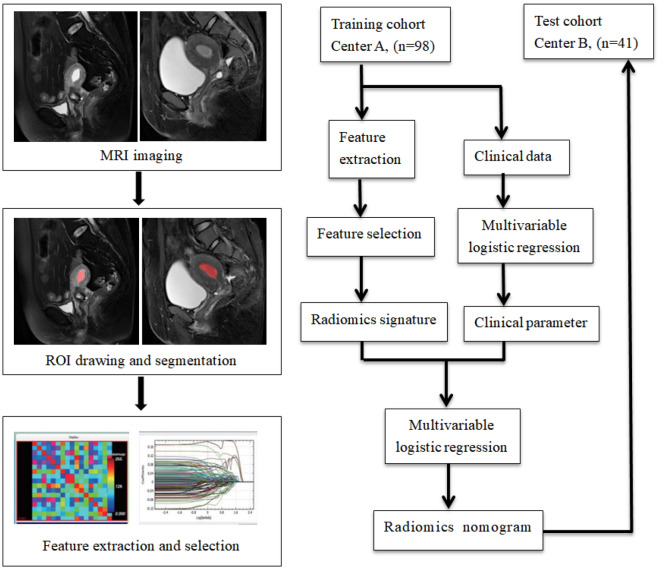
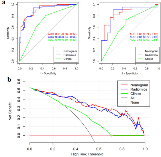
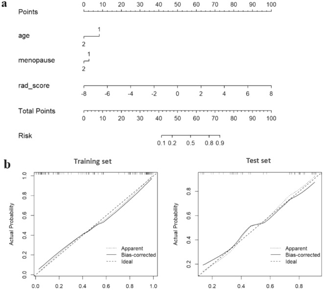
An unbiased and accurate diagnosis of benign and malignant endometrial lesions is essential for the gynecologist, as each type might require distinct treatment. Radiomics is a quantitative method that could facilitate deep mining of information and quantification of the heterogeneity in images, thereby aiding clinicians in proper lesion diagnosis. The aim of this study is to develop an appropriate predictive model for the classification of benign and malignant endometrial lesions, and evaluate potential clinical applicability of the model. 139 patients with pathologically-confirmed endometrial lesions from January 2018 to July 2020 in two independent centers (center A and B) were finally analyzed. Center A was used for training set, while center B was used for test set. The lesions were manually drawn on the largest slice based on the lesion area by two radiologists. After feature extraction and feature selection, the possible associations between radiomics features and clinical parameters were assessed by Uni- and multi- variable logistic regression. The receiver operator characteristic (ROC) curve and DeLong validation were employed to evaluate the possible predictive performance of the models. Decision curve analysis (DCA) was used to evaluate the net benefit of the radiomics nomogram. A radiomics prediction model was established from the 15 selected features, and were found to be relatively high discriminative on the basis of the area under the ROC curve (AUC) for both the training and the test cohorts (AUC = 0.90 and 0.85, respectively). The radiomics nomogram also showed good performance of discrimination for both the training and test cohorts (AUC = 0.91 and 0.86, respectively), and the DeLong test shows that AUCs were significantly different between clinical parameters and nomogram. The result of DCA demonstrated the clinical usefulness of this novel nomogram method. The predictive model constructed based on MRI radiomics and clinical parameters indicated a highly diagnostic efficiency, thereby implying its potential clinical usefulness for the precise identification and prediction of endometrial lesions.
对妇科医生而言,对良性和恶性子宫内膜病变进行公正和准确的诊断至关重要,因为每种类型可能需要不同的治疗方法。放射组学是一种定量方法,可以促进对信息的深度挖掘和对图像异质性的量化,从而帮助临床医生进行正确的病变诊断。本研究旨在为良性和恶性子宫内膜病变的分类建立一个合适的预测模型,并评估该模型的潜在临床适用性。最终分析了来自两个独立中心(中心 A 和 B)的 139 名经病理证实的子宫内膜病变患者。中心 A 用于训练集,中心 B 用于测试集。两位放射科医生根据病变面积在最大切片上手动绘制病变。在进行特征提取和特征选择后,通过单变量和多变量逻辑回归评估放射组学特征与临床参数之间的可能关联。采用接收者操作特征(ROC)曲线和 DeLong 验证评估模型的可能预测性能。决策曲线分析(DCA)用于评估放射组学列线图的净获益。基于 15 个选定特征建立了放射组学预测模型,基于 ROC 曲线下面积(AUC),该模型在训练和测试队列中均具有较高的区分能力(AUC 分别为 0.90 和 0.85)。放射组学列线图在训练和测试队列中也表现出良好的区分能力(AUC 分别为 0.91 和 0.86),DeLong 检验表明 AUC 在临床参数和列线图之间存在显著差异。DCA 的结果表明了这种新的列线图方法的临床实用性。基于 MRI 放射组学和临床参数构建的预测模型表明具有较高的诊断效率,这意味着其在子宫内膜病变的精确识别和预测方面具有潜在的临床应用价值。