Adhikari Surya Prasad, Paudel Astha, Sharma Anisha, Thapa Baruna, Khanal Neha, Shastri Nisha, Rai Sourav, Adhikari Rameshwar
Department of Mechanical and Aerospace Engineering, Institute of Engineering, Pulchowk Campus, Tribhuvan University, Lalitpur, Nepal.
College of Biomedical Engineering and Applied Sciences, Purbanchal University, Lalitpur, Nepal.
Int J Biomater. 2023 Jan 31;2023:8541621. doi: 10.1155/2023/8541621. eCollection 2023.
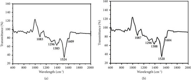
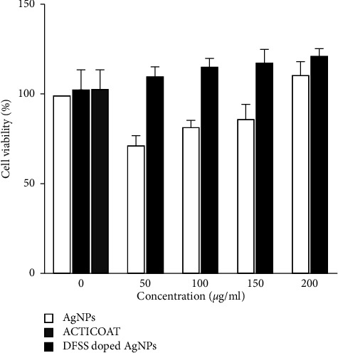
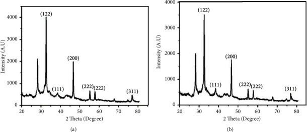
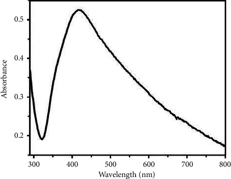
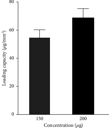
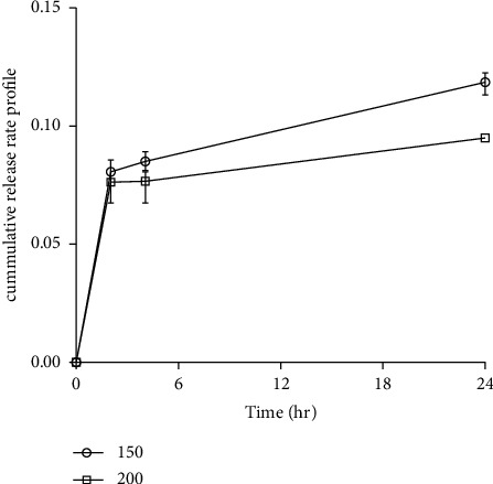
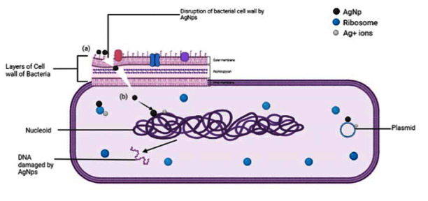
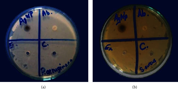
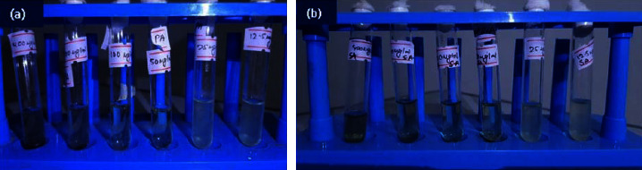
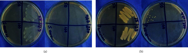
In this study, decellularized fish skin (DFS) scaffold decorated with silver nanoparticles was prepared for accelerating burn wound healing. The silver nanoparticles (AgNPs) synthesized by the green and facile method using leaf at different incubating times were characterized by using X-ray Diffraction (XRD), Fourier Transform Infrared (FT-IR) Spectroscopy, and Ultraviolet-Visible Spectroscopy (UV-Vis spectroscopy). The different characterizations confirmed that the sizes of AgNPs prepared by incubating for 6 hours and 12 hours were 29.1 nm and 35.2 nm, respectively. After that, the different concentrations of the smallest AgNPs were used to dope the DFS scaffold to determine the cell viability. Additionally, an agar well diffusion method was used to screen for antimicrobial activity. Minimum inhibitory concentration (MIC) and minimum bactericidal concentration (MBC) were used to correlate the concentration of AgNPs with its bactericidal effect which was seen from 50 g/ml. Then, the toxicity with human cells was investigated using a 3-(4, 5-dimethylthiazol-2-yl)-2, 5-diphenyl tetrazolium bromide (MTT) assay with no significant cell viability from the concentration of 50 g/ml to 200 g/ml compared to the cocultured and commercial treatments.
在本研究中,制备了用银纳米颗粒修饰的脱细胞鱼皮(DFS)支架,以加速烧伤创面愈合。使用X射线衍射(XRD)、傅里叶变换红外(FT-IR)光谱和紫外-可见光谱(UV-Vis光谱)对通过绿色简便方法使用树叶在不同孵育时间合成的银纳米颗粒(AgNPs)进行了表征。不同的表征证实,孵育6小时和12小时制备的AgNPs尺寸分别为29.1纳米和35.2纳米。之后,使用不同浓度的最小尺寸AgNPs对DFS支架进行掺杂,以确定细胞活力。此外,采用琼脂孔扩散法筛选抗菌活性。使用最低抑菌浓度(MIC)和最低杀菌浓度(MBC)来关联AgNPs的浓度与其杀菌效果,从50微克/毫升即可看出杀菌效果。然后,使用3-(4,5-二甲基噻唑-2-基)-2,5-二苯基溴化四氮唑(MTT)试验研究对人细胞的毒性,与共培养和商业处理相比,在50微克/毫升至2