Department of Ophthalmology, Visual and Anatomical Sciences, Wayne State University School of Medicine, Detroit, MI 48201, USA.
Department of Ophthalmology, Visual and Anatomical Sciences, Wayne State University School of Medicine, Detroit, MI 48201, USA.
STAR Protoc. 2023 Mar 17;4(1):102056. doi: 10.1016/j.xpro.2023.102056. Epub 2023 Jan 25.
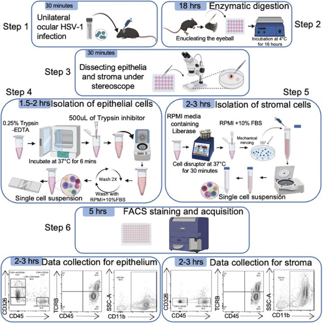
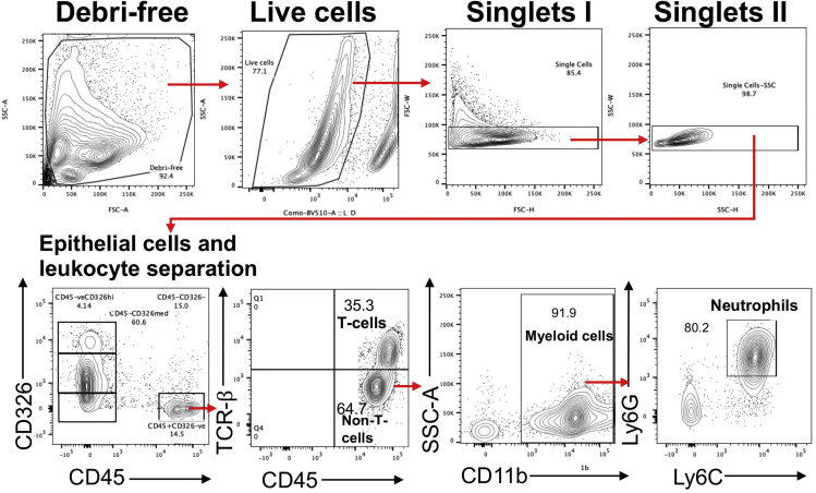
Existing flow cytometry approaches identify immune cells using the whole infected/inflamed cornea, which limits its ability to distinguish the immune cells infiltrating the corneal epithelium from the corneal stroma. Here, we present a protocol to analyze immune cells in the separated epithelium and stroma from naïve and herpes simplex virus-1 (HSV-1)-infected mouse corneas. We describe steps for viral infection, separation of corneal epithelium from stroma, preparation of a single-cell suspension of the individual epithelium and stroma, and flow cytometry assay.
现有的流式细胞术方法使用整个感染/炎症的角膜来识别免疫细胞,这限制了它区分浸润角膜上皮和基质的免疫细胞的能力。在这里,我们提出了一种从正常和单纯疱疹病毒 1(HSV-1)感染的小鼠角膜中分离的上皮和基质中分析免疫细胞的方案。我们描述了病毒感染、角膜上皮与基质分离、单个上皮和基质单细胞悬液制备以及流式细胞术检测的步骤。