• 文献检索
  • 文档翻译
  • 深度研究
  • 学术资讯
  • Suppr Zotero 插件Zotero 插件
  • 邀请有礼
  • 套餐&价格
  • 历史记录
应用&插件
Suppr Zotero 插件Zotero 插件浏览器插件Mac 客户端Windows 客户端微信小程序
定价
高级版会员购买积分包购买API积分包
服务
文献检索文档翻译深度研究API 文档MCP 服务
关于我们
关于 Suppr公司介绍联系我们用户协议隐私条款
关注我们

Suppr 超能文献

核心技术专利:CN118964589B侵权必究
粤ICP备2023148730 号-1Suppr @ 2026

文献检索

告别复杂PubMed语法,用中文像聊天一样搜索,搜遍4000万医学文献。AI智能推荐,让科研检索更轻松。

立即免费搜索

文件翻译

保留排版,准确专业,支持PDF/Word/PPT等文件格式,支持 12+语言互译。

免费翻译文档

深度研究

AI帮你快速写综述,25分钟生成高质量综述,智能提取关键信息,辅助科研写作。

立即免费体验

迈向基于临床的人类肠道细胞图谱通用坐标框架:肠道模型。

Towards a clinically-based common coordinate framework for the human gut cell atlas: the gut models.

机构信息

Department of Computer Science, School of Mathematical and Computer Sciences, Heriot-Watt University, Edinburgh, UK.

Division of Pathology, Centre for Comparative Pathology, Edinburgh Cancer Research Centre, Institute of Cancer and Genetics, University of Edinburgh, Crewe Road, Edinburgh, EH4 2XU, UK.

出版信息

BMC Med Inform Decis Mak. 2023 Feb 15;23(1):36. doi: 10.1186/s12911-023-02111-9.

DOI:10.1186/s12911-023-02111-9
PMID:36793076
原文链接:https://pmc.ncbi.nlm.nih.gov/articles/PMC9933383/
Abstract

BACKGROUND

The Human Cell Atlas resource will deliver single cell transcriptome data spatially organised in terms of gross anatomy, tissue location and with images of cellular histology. This will enable the application of bioinformatics analysis, machine learning and data mining revealing an atlas of cell types, sub-types, varying states and ultimately cellular changes related to disease conditions. To further develop the understanding of specific pathological and histopathological phenotypes with their spatial relationships and dependencies, a more sophisticated spatial descriptive framework is required to enable integration and analysis in spatial terms.

METHODS

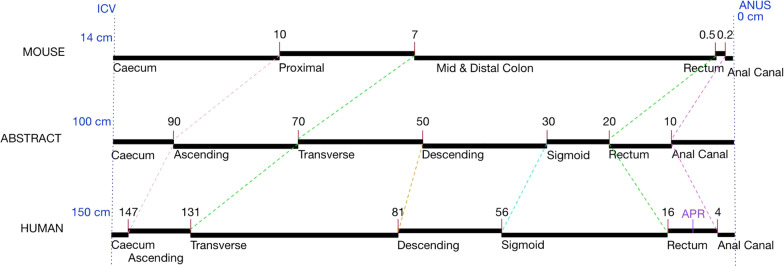
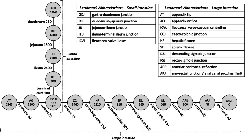
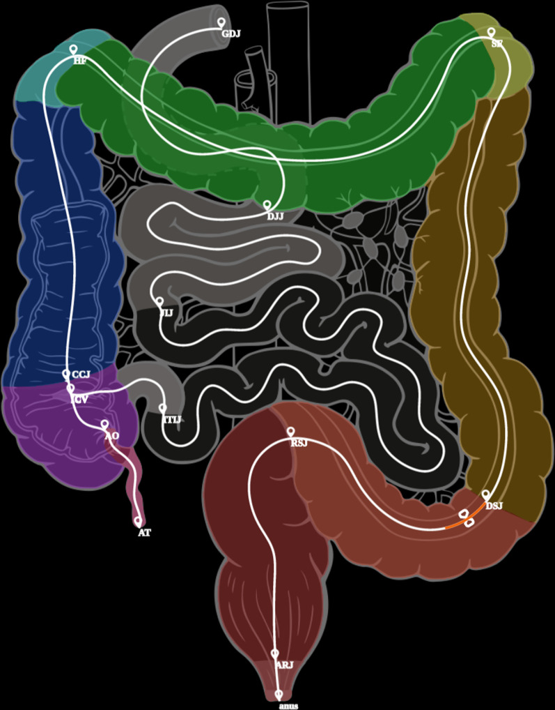
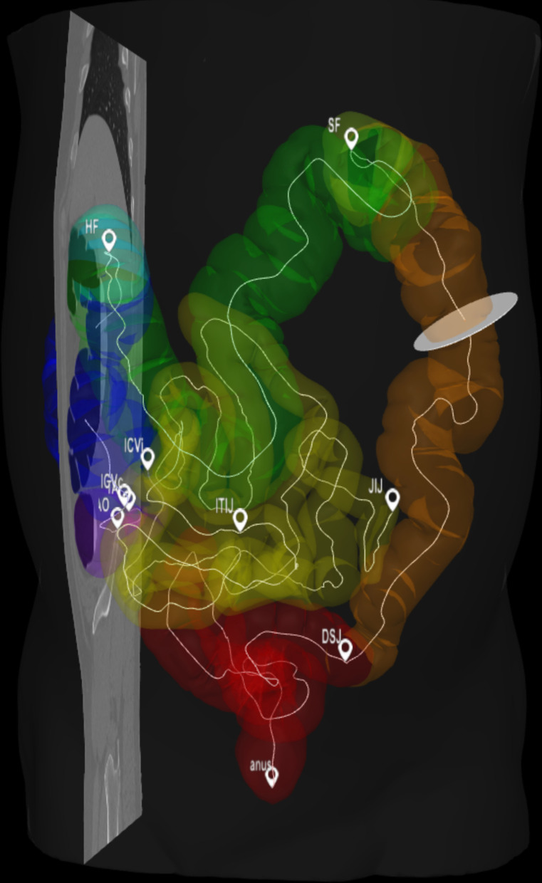
We describe a conceptual coordinate model for the Gut Cell Atlas (small and large intestines). Here, we focus on a Gut Linear Model (1-dimensional representation based on the centreline of the gut) that represents the location semantics as typically used by clinicians and pathologists when describing location in the gut. This knowledge representation is based on a set of standardised gut anatomy ontology terms describing regions in situ, such as ileum or transverse colon, and landmarks, such as ileo-caecal valve or hepatic flexure, together with relative or absolute distance measures. We show how locations in the 1D model can be mapped to and from points and regions in both a 2D model and 3D models, such as a patient's CT scan where the gut has been segmented.

RESULTS

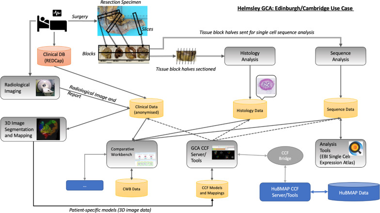
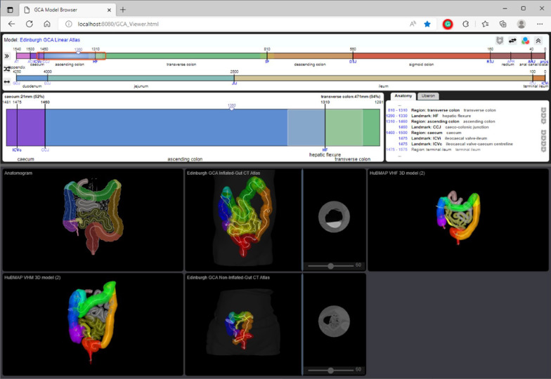
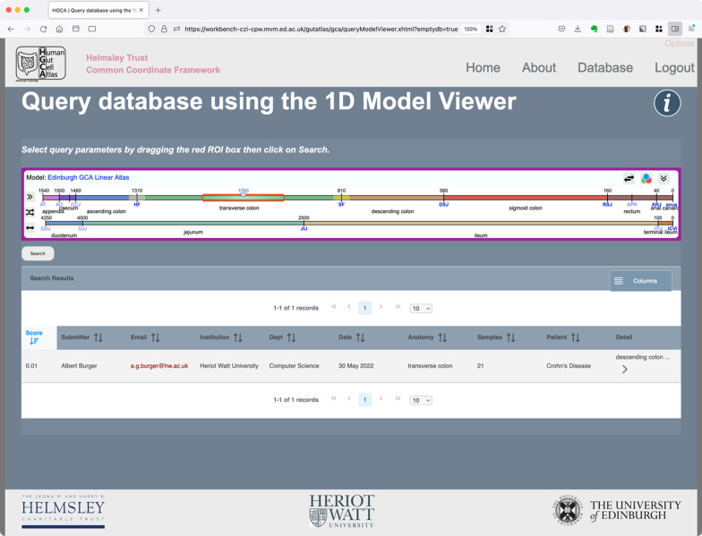
The outputs of this work include 1D, 2D and 3D models of the human gut, delivered through publicly accessible Json and image files. We also illustrate the mappings between models using a demonstrator tool that allows the user to explore the anatomical space of the gut. All data and software is fully open-source and available online.

CONCLUSIONS

Small and large intestines have a natural "gut coordinate" system best represented as a 1D centreline through the gut tube, reflecting functional differences. Such a 1D centreline model with landmarks, visualised using viewer software allows interoperable translation to both a 2D anatomogram model and multiple 3D models of the intestines. This permits users to accurately locate samples for data comparison.

摘要

背景

人类细胞图谱资源将提供根据大体解剖、组织位置以及细胞组织学图像组织的单细胞转录组数据。这将使生物信息学分析、机器学习和数据挖掘得以应用,揭示细胞类型、亚型、不同状态的图谱,并最终揭示与疾病状况相关的细胞变化。为了进一步了解具有空间关系和依赖性的特定病理和组织病理学表型,需要更复杂的空间描述框架来实现空间方面的集成和分析。

方法

我们描述了肠道细胞图谱(小肠和大肠)的概念坐标模型。在这里,我们专注于肠道线性模型(基于肠道中心线的一维表示),它代表了临床医生和病理学家在描述肠道位置时通常使用的位置语义。这种知识表示基于一组标准化的肠道解剖学本体术语,描述了原位区域,如回肠或横结肠,以及地标,如回盲瓣或肝曲,以及相对或绝对距离测量。我们展示了如何将一维模型中的位置映射到二维和三维模型(如肠道已分割的患者 CT 扫描)中的点和区域,以及如何将其反向映射。

结果

这项工作的输出包括通过可公开访问的 Json 和图像文件提供的一维、二维和三维人类肠道模型。我们还使用演示工具说明了模型之间的映射,该工具允许用户探索肠道的解剖空间。所有数据和软件均完全开源,并可在线获取。

结论

小肠和大肠具有天然的“肠道坐标”系统,最好用穿过肠道管的一维中心线表示,反映了功能差异。这种带有地标、使用查看器软件可视化的一维中心线模型可以与二维解剖图模型和多个肠道三维模型进行互操作转换。这允许用户准确定位样本以进行数据比较。

https://cdn.ncbi.nlm.nih.gov/pmc/blobs/217e/9933383/7688c3652709/12911_2023_2111_Fig8_HTML.jpg
https://cdn.ncbi.nlm.nih.gov/pmc/blobs/217e/9933383/7c2807de3b66/12911_2023_2111_Fig1_HTML.jpg
https://cdn.ncbi.nlm.nih.gov/pmc/blobs/217e/9933383/a57a3e8675a0/12911_2023_2111_Fig2_HTML.jpg
https://cdn.ncbi.nlm.nih.gov/pmc/blobs/217e/9933383/3f20d2199ab4/12911_2023_2111_Fig3_HTML.jpg
https://cdn.ncbi.nlm.nih.gov/pmc/blobs/217e/9933383/95af931771e8/12911_2023_2111_Fig4_HTML.jpg
https://cdn.ncbi.nlm.nih.gov/pmc/blobs/217e/9933383/e3f42c5bcc0e/12911_2023_2111_Fig5_HTML.jpg
https://cdn.ncbi.nlm.nih.gov/pmc/blobs/217e/9933383/f57008265ab2/12911_2023_2111_Fig6_HTML.jpg
https://cdn.ncbi.nlm.nih.gov/pmc/blobs/217e/9933383/264668b5512b/12911_2023_2111_Fig7_HTML.jpg
https://cdn.ncbi.nlm.nih.gov/pmc/blobs/217e/9933383/7688c3652709/12911_2023_2111_Fig8_HTML.jpg

相似文献

1
Towards a clinically-based common coordinate framework for the human gut cell atlas: the gut models.迈向基于临床的人类肠道细胞图谱通用坐标框架:肠道模型。
BMC Med Inform Decis Mak. 2023 Feb 15;23(1):36. doi: 10.1186/s12911-023-02111-9.
2
The NeuARt II system: a viewing tool for neuroanatomical data based on published neuroanatomical atlases.NeuARt II系统:一种基于已发表的神经解剖图谱的神经解剖数据查看工具。
BMC Bioinformatics. 2006 Dec 13;7:531. doi: 10.1186/1471-2105-7-531.
3
The HUDSEN Atlas: a three-dimensional (3D) spatial framework for studying gene expression in the developing human brain.HUDSEN 图谱:研究人类大脑发育过程中基因表达的三维(3D)空间框架。
J Anat. 2010 Oct;217(4):289-99. doi: 10.1111/j.1469-7580.2010.01290.x.
4
An analytical tool that quantifies cellular morphology changes from three-dimensional fluorescence images.一种可对三维荧光图像中的细胞形态变化进行量化的分析工具。
J Vis Exp. 2012 Aug 31(66):e4233. doi: 10.3791/4233.
5
Whole-Brain Image Analysis and Anatomical Atlas 3D Generation Using MagellanMapper.使用 MagellanMapper 进行全脑图像分析和解剖图谱 3D 生成。
Curr Protoc Neurosci. 2020 Dec;94(1):e104. doi: 10.1002/cpns.104.
6
Incorporating 3-dimensional models in online articles.将三维模型纳入在线文章中。
Am J Orthod Dentofacial Orthop. 2015 May;147(5 Suppl):S195-204. doi: 10.1016/j.ajodo.2015.02.002.
7
Constructing and optimizing 3D atlases from 2D data with application to the developing mouse brain.从二维数据构建和优化 3D 图谱及其在发育中老鼠大脑中的应用。
Elife. 2021 Feb 11;10:e61408. doi: 10.7554/eLife.61408.
8
NeuroTerrain--a client-server system for browsing 3D biomedical image data sets.NeuroTerrain——一个用于浏览三维生物医学图像数据集的客户端-服务器系统。
BMC Bioinformatics. 2007 Feb 5;8:40. doi: 10.1186/1471-2105-8-40.
9
A framework for representation and visualization of 3D shape variability of organs in an interactive anatomical atlas.一种用于在交互式解剖图谱中表示和可视化器官三维形状变异性的框架。
Methods Inf Med. 2009;48(3):272-81. doi: 10.3414/ME0551. Epub 2009 Mar 31.
10
A dynamic and expandable digital 3D-atlas maker for monitoring the temporal changes in tissue growth during hindbrain morphogenesis.用于监测后脑形态发生过程中组织生长的时间变化的动态可扩展数字 3D 图谱制作器。
Elife. 2022 Sep 28;11:e78300. doi: 10.7554/eLife.78300.

引用本文的文献

1
Single-cell and spatial transcriptomics of stricturing Crohn's disease highlights a fibrosis-associated network.狭窄性克罗恩病的单细胞和空间转录组学揭示了一个与纤维化相关的网络。
Nat Genet. 2025 Jun 25. doi: 10.1038/s41588-025-02225-y.
2
The human and non-human primate developmental GTEx projects.人类和非人类灵长类动物发育GTEx项目。
Nature. 2025 Jan;637(8046):557-564. doi: 10.1038/s41586-024-08244-9. Epub 2025 Jan 15.
3
Characterizing Low-cost Registration for Photographic Images to Computed Tomography.表征摄影图像到计算机断层扫描的低成本配准

本文引用的文献

1
Specimen, biological structure, and spatial ontologies in support of a Human Reference Atlas.支持人类参考图谱的样本、生物结构和空间本体论。
Sci Data. 2023 Mar 27;10(1):171. doi: 10.1038/s41597-023-01993-8.
2
Better, broader, safer: using health data for research and analysis (the Goldacre review).更好、更广泛、更安全:利用健康数据进行研究与分析(戈德acre报告)
J Radiol Prot. 2022 Sep 5;42(3). doi: 10.1088/1361-6498/ac89f8.
3
Single-cell RNA sequencing technologies and applications: A brief overview.单细胞 RNA 测序技术及应用:简述。
Proc SPIE Int Soc Opt Eng. 2024 Feb;12930. doi: 10.1117/12.3005578. Epub 2024 Apr 2.
4
The Comparative Pathology Workbench: Interactive visual analytics for biomedical data.比较病理学工作台:生物医学数据的交互式视觉分析
J Pathol Inform. 2023 Aug 9;14:100328. doi: 10.1016/j.jpi.2023.100328. eCollection 2023.
5
The Promise of Single-Cell RNA Sequencing to Redefine the Understanding of Crohn's Disease Fibrosis Mechanisms.单细胞RNA测序有望重新定义对克罗恩病纤维化机制的理解。
J Clin Med. 2023 Jun 7;12(12):3884. doi: 10.3390/jcm12123884.
6
A Roadmap for the Human Gut Cell Atlas.人类肠道细胞图谱研究路线图
Nat Rev Gastroenterol Hepatol. 2023 Sep;20(9):597-614. doi: 10.1038/s41575-023-00784-1. Epub 2023 May 31.
Clin Transl Med. 2022 Mar;12(3):e694. doi: 10.1002/ctm2.694.
4
FlyBase: a guided tour of highlighted features.FlyBase:特色功能导览
Genetics. 2022 Apr 4;220(4). doi: 10.1093/genetics/iyac035.
5
Zebrafish information network, the knowledgebase for Danio rerio research.斑马鱼信息网,斑马鱼研究知识库。
Genetics. 2022 Apr 4;220(4). doi: 10.1093/genetics/iyac016.
6
Expression Atlas update: gene and protein expression in multiple species.ExpressionAtlas 更新:多种物种中的基因和蛋白质表达。
Nucleic Acids Res. 2022 Jan 7;50(D1):D129-D140. doi: 10.1093/nar/gkab1030.
7
Anatomical structures, cell types and biomarkers of the Human Reference Atlas.人体参考图谱的解剖结构、细胞类型和生物标志物。
Nat Cell Biol. 2021 Nov;23(11):1117-1128. doi: 10.1038/s41556-021-00788-6. Epub 2021 Nov 8.
8
Multi-species single-cell transcriptomic analysis of ocular compartment regulons.眼内隔调控元的多物种单细胞转录组分析。
Nat Commun. 2021 Sep 28;12(1):5675. doi: 10.1038/s41467-021-25968-8.
9
Cells of the human intestinal tract mapped across space and time.人类肠道细胞的时空图谱。
Nature. 2021 Sep;597(7875):250-255. doi: 10.1038/s41586-021-03852-1. Epub 2021 Sep 8.
10
Regular Use of Proton Pump Inhibitor and the Risk of Inflammatory Bowel Disease: Pooled Analysis of 3 Prospective Cohorts.质子泵抑制剂的常规使用与炎症性肠病风险:3 项前瞻性队列的汇总分析。
Gastroenterology. 2021 Dec;161(6):1842-1852.e10. doi: 10.1053/j.gastro.2021.08.005. Epub 2021 Aug 11.