Woodland R M, El-Sheikh H, Darougar S, Squires S
J Clin Pathol. 1978 Nov;31(11):1073-7. doi: 10.1136/jcp.31.11.1073.
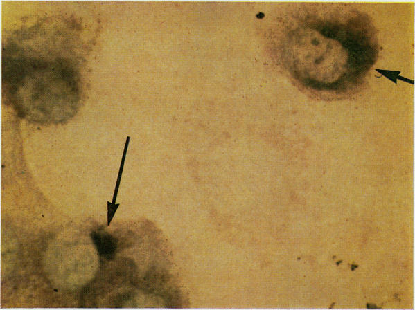
The sensitivities of Giemsa, immunofluorescence, and immunoperoxidase staining for the detection of Chlamydia psittaci inclusions in conjunctival scrapings and in irradiated McCoy cell monolayers were compared. Conjunctival specimens were obtained from a cat colony in which a trachoma-like disease, feline chlamydial keratoconjunctivitis, was endemic. The two immunochemical techniques were found to be of equal sensitivity and 50% to 100% more sensitive than Giemsa stain. Permanent preparations of immunoperoxidase stained material can be made and can be read using a simple light microscope. These features make the technique more useful than immunofluorescence staining, which gives temporary preparations that must be examined with a specialised fluorescence microscope.
比较了吉姆萨染色、免疫荧光和免疫过氧化物酶染色在检测结膜刮片和经辐照的 McCoy 细胞单层中鹦鹉热衣原体包涵体方面的敏感性。结膜标本取自一个猫群,其中一种类似沙眼的疾病——猫衣原体性角结膜炎呈地方性流行。发现这两种免疫化学技术具有相同的敏感性,且比吉姆萨染色敏感 50%至 100%。免疫过氧化物酶染色材料的永久制片可以制作,并且可以使用普通光学显微镜进行观察。这些特性使得该技术比免疫荧光染色更有用,免疫荧光染色得到的是临时制片,必须用专门的荧光显微镜进行检查。