• 文献检索
  • 文档翻译
  • 深度研究
  • 学术资讯
  • Suppr Zotero 插件Zotero 插件
  • 邀请有礼
  • 套餐&价格
  • 历史记录
应用&插件
Suppr Zotero 插件Zotero 插件浏览器插件Mac 客户端Windows 客户端微信小程序
定价
高级版会员购买积分包购买API积分包
服务
文献检索文档翻译深度研究API 文档MCP 服务
关于我们
关于 Suppr公司介绍联系我们用户协议隐私条款
关注我们

Suppr 超能文献

核心技术专利:CN118964589B侵权必究
粤ICP备2023148730 号-1Suppr @ 2026

文献检索

告别复杂PubMed语法,用中文像聊天一样搜索,搜遍4000万医学文献。AI智能推荐,让科研检索更轻松。

立即免费搜索

文件翻译

保留排版,准确专业,支持PDF/Word/PPT等文件格式,支持 12+语言互译。

免费翻译文档

深度研究

AI帮你快速写综述,25分钟生成高质量综述,智能提取关键信息,辅助科研写作。

立即免费体验

明场染色与荧光染色数据集——基于机器学习的虚拟染色的测试床图像集。

Brightfield vs Fluorescent Staining Dataset-A Test Bed Image Set for Machine Learning based Virtual Staining.

机构信息

Institute for Fundamental Medicine and Biology, Kazan Federal University, Kazan, 420008, Russia.

Centre for Digital Telecommunication Technologies, St. Petersburg Electrotechnical University "LETI", St. Petersburg, 197022, Russia.

出版信息

Sci Data. 2023 Mar 22;10(1):160. doi: 10.1038/s41597-023-02065-7.

DOI:10.1038/s41597-023-02065-7
PMID:36949058
原文链接:https://pmc.ncbi.nlm.nih.gov/articles/PMC10033900/
Abstract

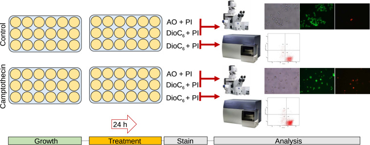
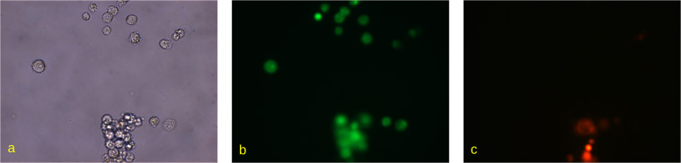
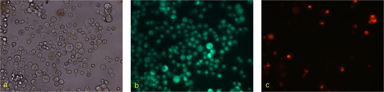
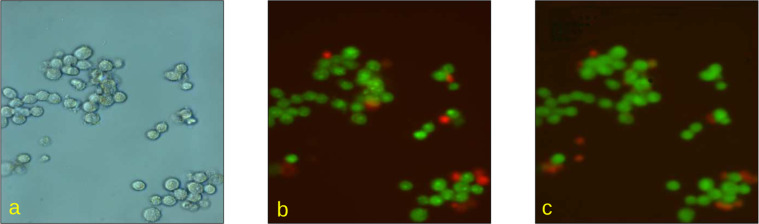
Differential fluorescent staining is an effective tool widely adopted for the visualization, segmentation and quantification of cells and cellular substructures as a part of standard microscopic imaging protocols. Incompatibility of staining agents with viable cells represents major and often inevitable limitations to its applicability in live experiments, requiring extraction of samples at different stages of experiment increasing laboratory costs. Accordingly, development of computerized image analysis methodology capable of segmentation and quantification of cells and cellular substructures from plain monochromatic images obtained by light microscopy without help of any physical markup techniques is of considerable interest. The enclosed set contains human colon adenocarcinoma Caco-2 cells microscopic images obtained under various imaging conditions with different viable vs non-viable cells fractions. Each field of view is provided in a three-fold representation, including phase-contrast microscopy and two differential fluorescent microscopy images with specific markup of viable and non-viable cells, respectively, produced using two different staining schemes, representing a prominent test bed for the validation of image analysis methods.

摘要

差示荧光染色是一种广泛应用于细胞和细胞亚结构可视化、分割和定量的有效工具,是标准显微镜成像方案的一部分。由于染色剂与活细胞不兼容,这代表着它在活体实验中的应用存在重大且往往不可避免的限制,需要在实验的不同阶段提取样本,从而增加了实验室成本。因此,开发能够对细胞和细胞亚结构进行分割和定量的计算机化图像分析方法,从没有任何物理标记技术帮助的普通单色显微镜图像中获取,这是非常有意义的。随函附上的是在不同成像条件下用不同死活细胞比例获得的人结肠腺癌 Caco-2 细胞的显微镜图像。每个视场提供了三重表示,包括相差显微镜和两种具有特定标记的差示荧光显微镜图像,分别使用两种不同的染色方案获得,这代表了用于验证图像分析方法的重要测试平台。

https://cdn.ncbi.nlm.nih.gov/pmc/blobs/4eb2/10033900/0b18187c221c/41597_2023_2065_Fig5_HTML.jpg
https://cdn.ncbi.nlm.nih.gov/pmc/blobs/4eb2/10033900/ea93dc96bd25/41597_2023_2065_Fig1_HTML.jpg
https://cdn.ncbi.nlm.nih.gov/pmc/blobs/4eb2/10033900/8dbb2d1e2564/41597_2023_2065_Fig2_HTML.jpg
https://cdn.ncbi.nlm.nih.gov/pmc/blobs/4eb2/10033900/68d734b121c0/41597_2023_2065_Fig3_HTML.jpg
https://cdn.ncbi.nlm.nih.gov/pmc/blobs/4eb2/10033900/9f8b6d8f1b50/41597_2023_2065_Fig4_HTML.jpg
https://cdn.ncbi.nlm.nih.gov/pmc/blobs/4eb2/10033900/0b18187c221c/41597_2023_2065_Fig5_HTML.jpg

相似文献

1
Brightfield vs Fluorescent Staining Dataset-A Test Bed Image Set for Machine Learning based Virtual Staining.明场染色与荧光染色数据集——基于机器学习的虚拟染色的测试床图像集。
Sci Data. 2023 Mar 22;10(1):160. doi: 10.1038/s41597-023-02065-7.
2
Weakly Supervised Learning using Attention gates for colon cancer histopathological image segmentation.基于注意力门控的弱监督学习在结肠癌病理图像分割中的应用。
Artif Intell Med. 2022 Nov;133:102407. doi: 10.1016/j.artmed.2022.102407. Epub 2022 Sep 24.
3
AutoIHC-Analyzer: computer-assisted microscopy for automated membrane extraction/scoring in HER2 molecular markers.AutoIHC-Analyzer:用于 HER2 分子标志物的自动膜提取/评分的计算机辅助显微镜。
J Microsc. 2021 Jan;281(1):87-96. doi: 10.1111/jmi.12955. Epub 2020 Aug 27.
4
A Dataset and a Technique for Generalized Nuclear Segmentation for Computational Pathology.用于计算病理学中通用核分割的数据集和技术。
IEEE Trans Med Imaging. 2017 Jul;36(7):1550-1560. doi: 10.1109/TMI.2017.2677499. Epub 2017 Mar 6.
5
Suggestive annotation of brain MR images with gradient-guided sampling.基于梯度引导采样的脑磁共振图像提示性标注。
Med Image Anal. 2022 Apr;77:102373. doi: 10.1016/j.media.2022.102373. Epub 2022 Jan 24.
6
Large scale tissue histopathology image classification, segmentation, and visualization via deep convolutional activation features.通过深度卷积激活特征进行大规模组织病理图像分类、分割和可视化
BMC Bioinformatics. 2017 May 26;18(1):281. doi: 10.1186/s12859-017-1685-x.
7
HCS Methodology for Helping in Lab Scale Image-Based Assays.用于实验室规模基于图像分析的帮助的HCS方法学。
Methods Mol Biol. 2019;2040:331-356. doi: 10.1007/978-1-4939-9686-5_15.
8
Learning-based 3T brain MRI segmentation with guidance from 7T MRI labeling.基于学习的3T脑磁共振成像分割,由7T磁共振成像标记引导。
Med Phys. 2016 Dec;43(12):6588-6597. doi: 10.1118/1.4967487.
9
Practical segmentation of nuclei in brightfield cell images with neural networks trained on fluorescently labelled samples.基于荧光标记样本训练的神经网络对明场细胞图像中的细胞核进行实用分割。
J Microsc. 2021 Oct;284(1):12-24. doi: 10.1111/jmi.13038. Epub 2021 Jun 15.
10
Protocols for staining of bile canalicular and sinusoidal networks of human, mouse and pig livers, three-dimensional reconstruction and quantification of tissue microarchitecture by image processing and analysis.人、小鼠和猪肝脏胆小管和肝血窦网络染色方案、通过图像处理和分析进行组织微结构的三维重建和定量分析。
Arch Toxicol. 2014 May;88(5):1161-83. doi: 10.1007/s00204-014-1243-5. Epub 2014 Apr 19.

引用本文的文献

1
Instance segmentation of cells and nuclei from multi-organ cross-protocol microscopic images.从多器官跨协议显微图像中对细胞和细胞核进行实例分割。
Quant Imaging Med Surg. 2024 Sep 1;14(9):6204-6221. doi: 10.21037/qims-24-801. Epub 2024 Aug 28.

本文引用的文献

1
LIVECell-A large-scale dataset for label-free live cell segmentation.LiveCell-A 大型无标记活细胞分割数据集。
Nat Methods. 2021 Sep;18(9):1038-1045. doi: 10.1038/s41592-021-01249-6. Epub 2021 Aug 30.
2
EVICAN-a balanced dataset for algorithm development in cell and nucleus segmentation.EVICAN-用于细胞和细胞核分割算法开发的平衡数据集。
Bioinformatics. 2020 Jun 1;36(12):3863-3870. doi: 10.1093/bioinformatics/btaa225.
3
The Quantitative-Phase Dynamics of Apoptosis and Lytic Cell Death.细胞凋亡和溶细胞性细胞死亡的定量相动力学。
Sci Rep. 2020 Jan 31;10(1):1566. doi: 10.1038/s41598-020-58474-w.
4
Machine learning approach of automatic identification and counting of blood cells.血细胞自动识别与计数的机器学习方法。
Healthc Technol Lett. 2019 Jul 17;6(4):103-108. doi: 10.1049/htl.2018.5098. eCollection 2019 Aug.
5
Fast and simple tool for the quantification of biofilm-embedded cells sub-populations from fluorescent microscopic images.一种快速简单的工具,可从荧光显微镜图像中定量生物膜嵌入细胞的亚群。
PLoS One. 2018 May 1;13(5):e0193267. doi: 10.1371/journal.pone.0193267. eCollection 2018.
6
Annotated high-throughput microscopy image sets for validation.用于验证的带注释的高通量显微镜图像集。
Nat Methods. 2012 Jun 28;9(7):637. doi: 10.1038/nmeth.2083.