• 文献检索
  • 文档翻译
  • 深度研究
  • 学术资讯
  • Suppr Zotero 插件Zotero 插件
  • 邀请有礼
  • 套餐&价格
  • 历史记录
应用&插件
Suppr Zotero 插件Zotero 插件浏览器插件Mac 客户端Windows 客户端微信小程序
定价
高级版会员购买积分包购买API积分包
服务
文献检索文档翻译深度研究API 文档MCP 服务
关于我们
关于 Suppr公司介绍联系我们用户协议隐私条款
关注我们

Suppr 超能文献

核心技术专利:CN118964589B侵权必究
粤ICP备2023148730 号-1Suppr @ 2026

文献检索

告别复杂PubMed语法,用中文像聊天一样搜索,搜遍4000万医学文献。AI智能推荐,让科研检索更轻松。

立即免费搜索

文件翻译

保留排版,准确专业,支持PDF/Word/PPT等文件格式,支持 12+语言互译。

免费翻译文档

深度研究

AI帮你快速写综述,25分钟生成高质量综述,智能提取关键信息,辅助科研写作。

立即免费体验

基于epoch 和准确率的深度学习技术在心脏 MRI 分割中的实证研究。

Epoch and accuracy based empirical study for cardiac MRI segmentation using deep learning technique.

机构信息

Maulana Azad National Institute of Technology, Bhopal, India.

出版信息

PeerJ. 2023 Mar 22;11:e14939. doi: 10.7717/peerj.14939. eCollection 2023.

DOI:10.7717/peerj.14939
PMID:36974136
原文链接:https://pmc.ncbi.nlm.nih.gov/articles/PMC10039650/
Abstract

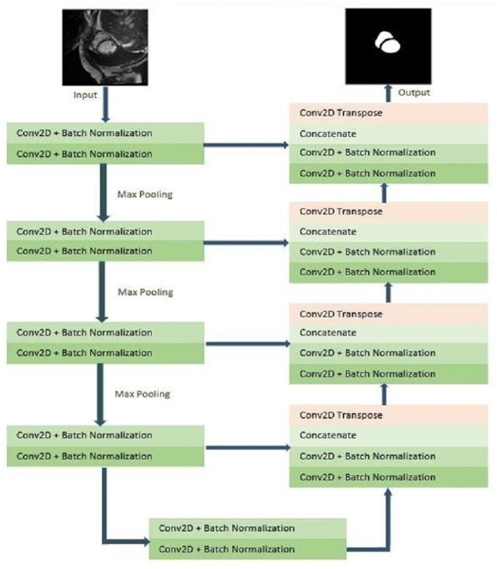
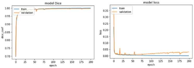
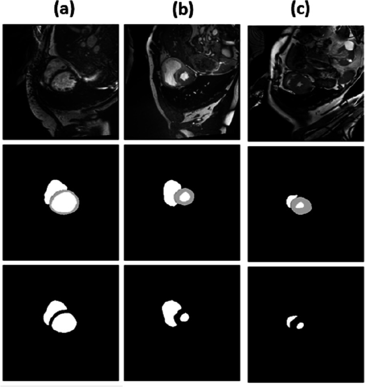
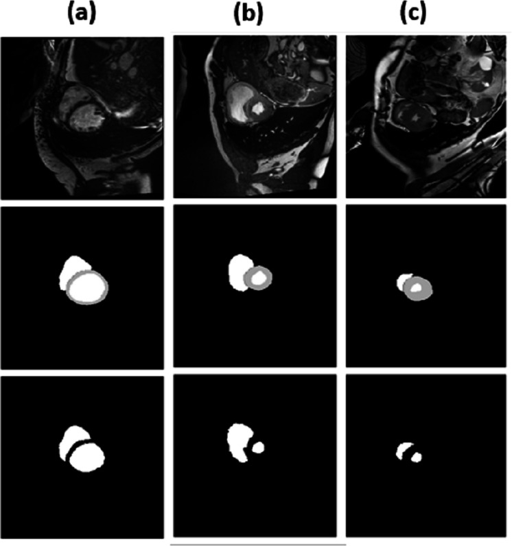
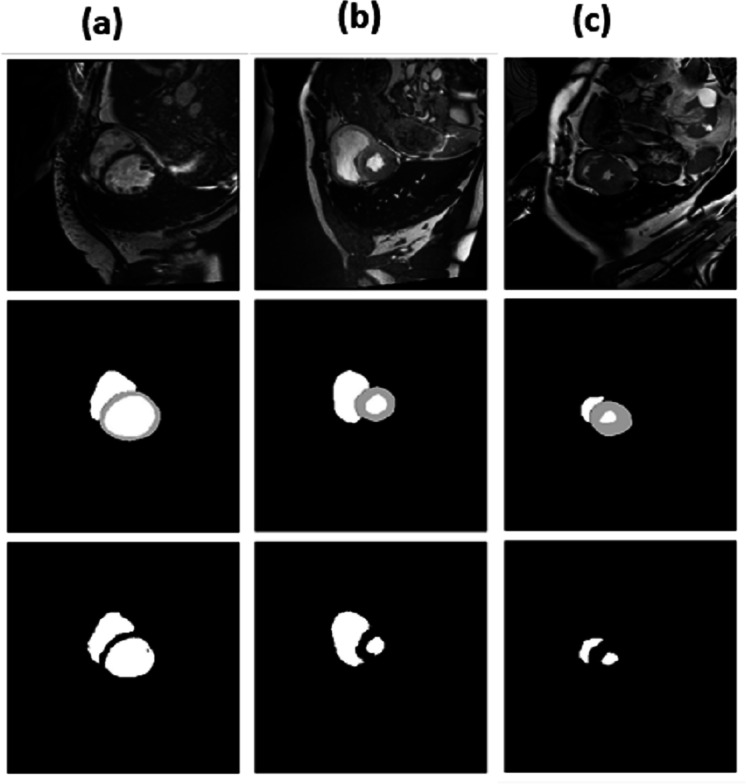
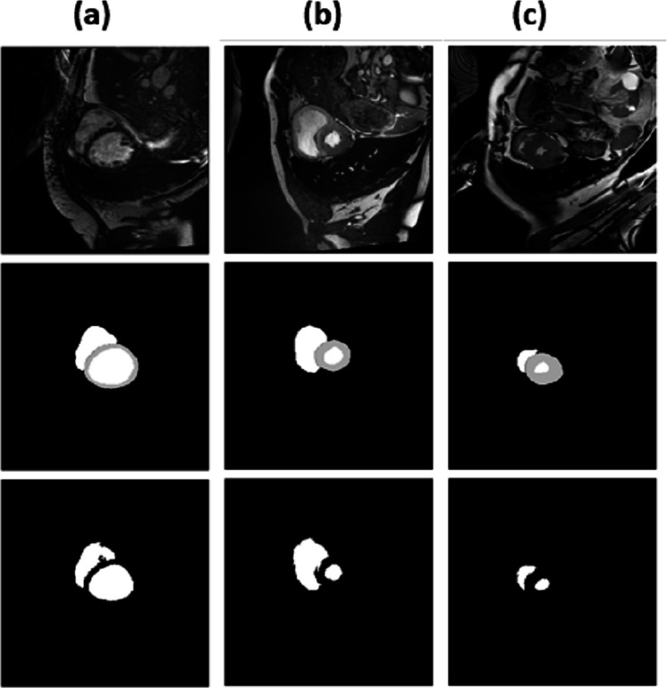
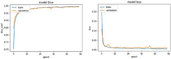
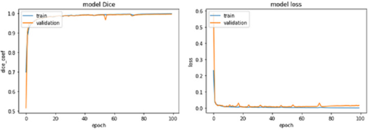
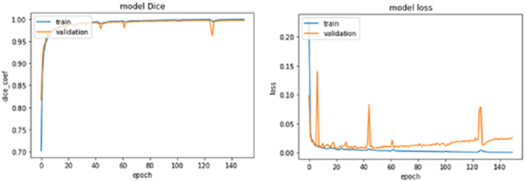
Cardiac magnetic resonance imaging (CMRI) is a non-invasive imaging technique to analyse the structure and function of the heart. It was enhanced considerably over several years to deliver functional information for diagnosing and managing cardiovascular disease. CMRI image delivers non-invasive, clear access to the heart and great vessels. The segmentation of CMRI provides quantification parameters such as myocardial viability, ejection fraction, cardiac chamber volume, and morphological details. In general, experts interpret the CMR images by delineating the images manually. The manual segmentation process is time-consuming, and it has been observed that the final observation varied with the opinion of the different experts. Convolution neural network is a new-age technology that provides impressive results compared to manual ones. In this study convolution neural network model is used for the segmentation task. The neural network parameters have been optimized to perform on the novel data set for accurate predictions. With other parameters, epochs play an essential role in training the network, as the network should not be under-fitted or over-fitted. The relationship between the hyperparameter epoch and accuracy is established in the model. The model delivers the accuracy of 0.88 in terms of the IoU coefficient.

摘要

心脏磁共振成像(CMRI)是一种非侵入性成像技术,用于分析心脏的结构和功能。它在过去几年中得到了极大的增强,能够提供用于诊断和治疗心血管疾病的功能信息。CMRI 图像提供了对心脏和大血管的非侵入性、清晰的访问。CMRI 的分割提供了心肌活力、射血分数、心脏腔室容积和形态细节等定量参数。通常,专家通过手动描绘图像来解释 CMR 图像。手动分割过程非常耗时,并且已经观察到最终观察结果因不同专家的意见而有所不同。卷积神经网络是一种新技术,与手动方法相比,它提供了令人印象深刻的结果。在这项研究中,卷积神经网络模型用于分割任务。神经网络参数经过优化,可在新的数据集上进行准确预测。在训练网络时,其他参数中,时期起着至关重要的作用,因为网络既不能欠拟合也不能过拟合。在模型中建立了超参数时期与准确性之间的关系。该模型的 IoU 系数准确率为 0.88。

https://cdn.ncbi.nlm.nih.gov/pmc/blobs/6216/10039650/44a7ffd1cf12/peerj-11-14939-g009.jpg
https://cdn.ncbi.nlm.nih.gov/pmc/blobs/6216/10039650/28201b37d03f/peerj-11-14939-g001.jpg
https://cdn.ncbi.nlm.nih.gov/pmc/blobs/6216/10039650/30e7005b38df/peerj-11-14939-g002.jpg
https://cdn.ncbi.nlm.nih.gov/pmc/blobs/6216/10039650/0ba673d65786/peerj-11-14939-g003.jpg
https://cdn.ncbi.nlm.nih.gov/pmc/blobs/6216/10039650/e49efb90c4b0/peerj-11-14939-g004.jpg
https://cdn.ncbi.nlm.nih.gov/pmc/blobs/6216/10039650/de8f275cce1d/peerj-11-14939-g005.jpg
https://cdn.ncbi.nlm.nih.gov/pmc/blobs/6216/10039650/09cb0f0bff83/peerj-11-14939-g006.jpg
https://cdn.ncbi.nlm.nih.gov/pmc/blobs/6216/10039650/ce1b5864a225/peerj-11-14939-g007.jpg
https://cdn.ncbi.nlm.nih.gov/pmc/blobs/6216/10039650/e82a6422ba86/peerj-11-14939-g008.jpg
https://cdn.ncbi.nlm.nih.gov/pmc/blobs/6216/10039650/44a7ffd1cf12/peerj-11-14939-g009.jpg

相似文献

1
Epoch and accuracy based empirical study for cardiac MRI segmentation using deep learning technique.基于epoch 和准确率的深度学习技术在心脏 MRI 分割中的实证研究。
PeerJ. 2023 Mar 22;11:e14939. doi: 10.7717/peerj.14939. eCollection 2023.
2
Magnetic Resonance Imaging Images Based Brain Tumor Extraction, Segmentation and Detection Using Convolutional Neural Network and VGC 16 Model.基于卷积神经网络和 VGC16 模型的磁共振成像图像脑肿瘤提取、分割与检测
Am J Clin Oncol. 2024 Jul 1;47(7):339-349. doi: 10.1097/COC.0000000000001097. Epub 2024 Apr 16.
3
CardSegNet: An adaptive hybrid CNN-vision transformer model for heart region segmentation in cardiac MRI.CardSegNet:一种自适应混合 CNN-vision 变压器模型,用于心脏 MRI 中的心脏区域分割。
Comput Med Imaging Graph. 2024 Jul;115:102382. doi: 10.1016/j.compmedimag.2024.102382. Epub 2024 Apr 16.
4
Fully automated cardiac MRI segmentation using dilated residual network.使用扩张残差网络的全自动心脏磁共振成像分割
Med Phys. 2023 Apr;50(4):2162-2175. doi: 10.1002/mp.16108. Epub 2022 Dec 7.
5
Automatic left ventricle segmentation from cardiac magnetic resonance images using a capsule network.基于胶囊网络的心脏磁共振图像左心室自动分割。
J Xray Sci Technol. 2020;28(3):541-553. doi: 10.3233/XST-190621.
6
Medical image diagnosis of prostate tumor based on PSP-Net+VGG16 deep learning network.基于 PSP-Net+VGG16 深度学习网络的前列腺肿瘤医学影像诊断。
Comput Methods Programs Biomed. 2022 Jun;221:106770. doi: 10.1016/j.cmpb.2022.106770. Epub 2022 Mar 23.
7
Automated left ventricular segmentation from cardiac magnetic resonance images via adversarial learning with multi-stage pose estimation network and co-discriminator.基于多阶段姿态估计网络和协同鉴别器的对抗学习实现心脏磁共振图像左心室自动分割。
Med Image Anal. 2021 Feb;68:101891. doi: 10.1016/j.media.2020.101891. Epub 2020 Nov 11.
8
Deeply supervised 3D fully convolutional networks with group dilated convolution for automatic MRI prostate segmentation.基于深度监督的三维全卷积网络与分组空洞卷积在自动 MRI 前列腺分割中的应用。
Med Phys. 2019 Apr;46(4):1707-1718. doi: 10.1002/mp.13416. Epub 2019 Feb 19.
9
A distance map regularized CNN for cardiac cine MR image segmentation.基于距离图正则化卷积神经网络的心电影磁共振图像分割。
Med Phys. 2019 Dec;46(12):5637-5651. doi: 10.1002/mp.13853. Epub 2019 Oct 31.
10
Cardiac MR segmentation based on sequence propagation by deep learning.基于深度学习的序列传播的心脏磁共振分割。
PLoS One. 2020 Apr 9;15(4):e0230415. doi: 10.1371/journal.pone.0230415. eCollection 2020.

引用本文的文献

1
Accelerated Cardiac MRI with Deep Learning-based Image Reconstruction for Cine Imaging.基于深度学习的图像重建的加速心脏 MRI 用于电影成像。
2
Encoder-decoder convolutional neural network for simple CT segmentation of COVID-19 infected lungs.用于COVID-19感染肺部简单CT分割的编码器-解码器卷积神经网络。
PeerJ Comput Sci. 2024 Jul 23;10:e2178. doi: 10.7717/peerj-cs.2178. eCollection 2024.

本文引用的文献

1
Bridging 2D and 3D segmentation networks for computation-efficient volumetric medical image segmentation: An empirical study of 2.5D solutions.桥接二维和三维分割网络以实现计算高效的容积医学图像分割:对 2.5D 解决方案的实证研究。
Comput Med Imaging Graph. 2022 Jul;99:102088. doi: 10.1016/j.compmedimag.2022.102088. Epub 2022 Jun 9.
2
Impact of Image Resolution on Deep Learning Performance in Endoscopy Image Classification: An Experimental Study Using a Large Dataset of Endoscopic Images.图像分辨率对内镜图像分类中深度学习性能的影响:使用大型内镜图像数据集的实验研究
Diagnostics (Basel). 2021 Nov 24;11(12):2183. doi: 10.3390/diagnostics11122183.
3
Deep neural network ensemble for on-the-fly quality control-driven segmentation of cardiac MRI T1 mapping.
基于深度神经网络集成的心脏 MRI T1 映射实时质量控制驱动分割。
Med Image Anal. 2021 Jul;71:102029. doi: 10.1016/j.media.2021.102029. Epub 2021 Mar 11.
4
Automated cardiovascular magnetic resonance image analysis with fully convolutional networks.基于全卷积网络的自动化心血管磁共振图像分析。
J Cardiovasc Magn Reson. 2018 Sep 14;20(1):65. doi: 10.1186/s12968-018-0471-x.
5
Deep Learning Techniques for Automatic MRI Cardiac Multi-Structures Segmentation and Diagnosis: Is the Problem Solved?深度学习技术在自动 MRI 心脏多结构分割与诊断中的应用:问题是否已解决?
IEEE Trans Med Imaging. 2018 Nov;37(11):2514-2525. doi: 10.1109/TMI.2018.2837502. Epub 2018 May 17.
6
Segmentation of Left and Right Ventricles in Cardiac MRI Using Active Contours.基于活动轮廓模型的心脏磁共振成像中左右心室分割
Comput Math Methods Med. 2017;2017:8350680. doi: 10.1155/2017/8350680. Epub 2017 Aug 8.
7
A combined deep-learning and deformable-model approach to fully automatic segmentation of the left ventricle in cardiac MRI.一种联合深度学习和可变形模型的方法,用于心脏 MRI 中左心室的全自动分割。
Med Image Anal. 2016 May;30:108-119. doi: 10.1016/j.media.2016.01.005. Epub 2016 Feb 6.