• 文献检索
  • 文档翻译
  • 深度研究
  • 学术资讯
  • Suppr Zotero 插件Zotero 插件
  • 邀请有礼
  • 套餐&价格
  • 历史记录
应用&插件
Suppr Zotero 插件Zotero 插件浏览器插件Mac 客户端Windows 客户端微信小程序
定价
高级版会员购买积分包购买API积分包
服务
文献检索文档翻译深度研究API 文档MCP 服务
关于我们
关于 Suppr公司介绍联系我们用户协议隐私条款
关注我们

Suppr 超能文献

核心技术专利:CN118964589B侵权必究
粤ICP备2023148730 号-1Suppr @ 2026

文献检索

告别复杂PubMed语法,用中文像聊天一样搜索,搜遍4000万医学文献。AI智能推荐,让科研检索更轻松。

立即免费搜索

文件翻译

保留排版,准确专业,支持PDF/Word/PPT等文件格式,支持 12+语言互译。

免费翻译文档

深度研究

AI帮你快速写综述,25分钟生成高质量综述,智能提取关键信息,辅助科研写作。

立即免费体验

使用机器学习检测致痫性病变中的皮质厚度变化

Detecting Cortical Thickness Changes in Epileptogenic Lesions Using Machine Learning.

作者信息

Azzony Sumayya, Moria Kawthar, Alghamdi Jamaan

机构信息

Department of Computer Sciences, Faculty of Computing and Information Technology, King Abdulaziz University, Jeddah 21589, Saudi Arabia.

Diagnostic Radiology Department, Faculty of Applied Medical Sciences, King Abdulaziz University, Jeddah 21589, Saudi Arabia.

出版信息

Brain Sci. 2023 Mar 14;13(3):487. doi: 10.3390/brainsci13030487.

DOI:10.3390/brainsci13030487
PMID:36979297
原文链接:https://pmc.ncbi.nlm.nih.gov/articles/PMC10046408/
Abstract

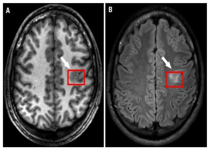
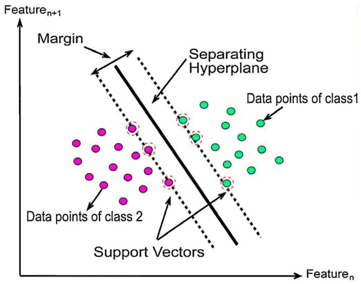
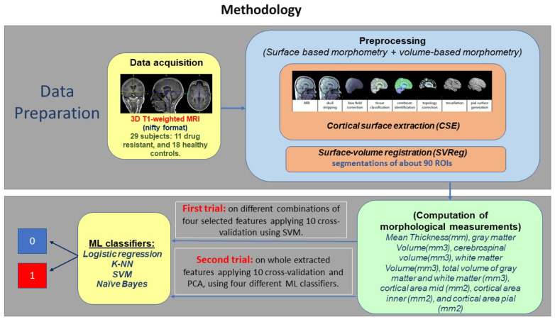
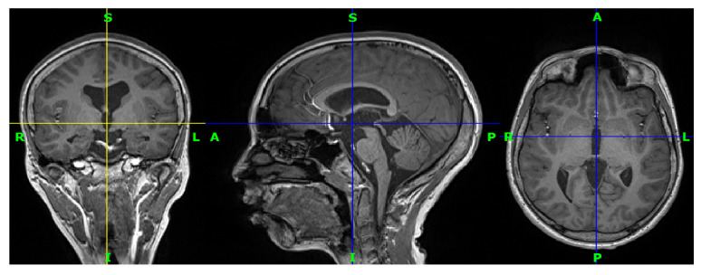
Epilepsy is a neurological disorder characterized by abnormal brain activity. Epileptic patients suffer from unpredictable seizures, which may cause a loss of awareness. Seizures are considered drug resistant if treatment does not affect success. This leads practitioners to calculate the cortical thickness to measure the distance between the brain's white and grey matter surfaces at various locations to perform a surgical intervention. In this study, we introduce using machine learning as an approach to classify extracted measurements from T1-weighted magnetic resonance imaging. Data were collected from the epilepsy unit at King Abdulaziz University Hospital. We applied two trials to classify the extracted measurements from T1-weighted MRI for drug-resistant epilepsy and healthy control subjects. The preprocessing sequence on T1-weighted MRI images was performed using C++ through BrainSuite's pipeline. The first trial was performed on seven different combinations of four commonly selected measurements. The best performance was achieved in Exp6 and Exp7, with 80.00% accuracy, 83.00% recall score, and 83.88% precision. It is noticeable that grey matter volume and white matter volume measurements are more significant than the cortical thickness measurement. The second trial applied four different machine learning classifiers after applying 10-fold cross-validation and principal component analysis on all extracted measurements as in the first trial based on the mentioned previous works. The K-nearest neighbours model outperformed the other machine learning classifiers with 97.11% accuracy, 75.00% recall score, and 75.00% precision.

摘要

癫痫是一种以大脑异常活动为特征的神经系统疾病。癫痫患者会遭受不可预测的发作,这可能导致意识丧失。如果治疗无效,发作则被认为是耐药性的。这使得从业者计算皮质厚度,以测量大脑不同位置白质和灰质表面之间的距离,从而进行手术干预。在本研究中,我们引入使用机器学习作为一种方法,对从T1加权磁共振成像中提取的测量数据进行分类。数据是从阿卜杜勒阿齐兹国王大学医院的癫痫科收集的。我们应用两种试验,对从T1加权MRI中提取的测量数据进行分类,以区分耐药性癫痫患者和健康对照者。使用C++通过BrainSuite的管道对T1加权MRI图像执行预处理序列。第一次试验在四个常用测量值的七种不同组合上进行。在Exp6和Exp7中取得了最佳性能,准确率为80.00%,召回率为83.00%,精确率为83.88%。值得注意的是,灰质体积和白质体积测量比皮质厚度测量更显著。第二次试验在如第一次试验那样对所有提取的测量数据应用10倍交叉验证和主成分分析之后,应用了四种不同的机器学习分类器。K近邻模型的表现优于其他机器学习分类器,准确率为97.11%,召回率为75.00%,精确率为75.00%。

https://cdn.ncbi.nlm.nih.gov/pmc/blobs/7420/10046408/9d20c85a25f0/brainsci-13-00487-g006.jpg
https://cdn.ncbi.nlm.nih.gov/pmc/blobs/7420/10046408/436ec8935f5f/brainsci-13-00487-g001.jpg
https://cdn.ncbi.nlm.nih.gov/pmc/blobs/7420/10046408/fbc027b37b22/brainsci-13-00487-g003.jpg
https://cdn.ncbi.nlm.nih.gov/pmc/blobs/7420/10046408/fcadc658c1de/brainsci-13-00487-g004.jpg
https://cdn.ncbi.nlm.nih.gov/pmc/blobs/7420/10046408/d0e74db4bcc6/brainsci-13-00487-g005.jpg
https://cdn.ncbi.nlm.nih.gov/pmc/blobs/7420/10046408/9d20c85a25f0/brainsci-13-00487-g006.jpg

相似文献

1
Detecting Cortical Thickness Changes in Epileptogenic Lesions Using Machine Learning.使用机器学习检测致痫性病变中的皮质厚度变化
Brain Sci. 2023 Mar 14;13(3):487. doi: 10.3390/brainsci13030487.
2
Analysis of structural brain MRI and multi-parameter classification for Alzheimer's disease.阿尔茨海默病的脑结构磁共振成像分析及多参数分类
Biomed Tech (Berl). 2018 Jul 26;63(4):427-437. doi: 10.1515/bmt-2016-0239.
3
Gray-matter-specific MR imaging improves the detection of epileptogenic zones in focal cortical dysplasia: A new sequence called fluid and white matter suppression (FLAWS).灰质特异性磁共振成像提高局灶性皮质发育不良致痫灶的检出:一种新的序列,称为液体和白质抑制(FLAWS)。
Neuroimage Clin. 2018 Aug 11;20:388-397. doi: 10.1016/j.nicl.2018.08.010. eCollection 2018.
4
Surface-based laminar analysis of diffusion abnormalities in cortical and white matter layers in neocortical epilepsy.脑皮质癫痫中皮质和白质各层弥散异常的基于表面的层分析。
Epilepsia. 2013 Apr;54(4):667-77. doi: 10.1111/epi.12129. Epub 2013 Feb 28.
5
Computational analysis in epilepsy neuroimaging: A survey of features and methods.癫痫神经影像学中的计算分析:特征与方法综述
Neuroimage Clin. 2016 Feb 23;11:515-529. doi: 10.1016/j.nicl.2016.02.013. eCollection 2016.
6
Epileptic Patient Activity Recognition System Using Extreme Learning Machine Method.基于极限学习机方法的癫痫患者活动识别系统
Biomedicines. 2023 Mar 7;11(3):816. doi: 10.3390/biomedicines11030816.
7
Cortical feature analysis and machine learning improves detection of "MRI-negative" focal cortical dysplasia.皮质特征分析和机器学习可提高对“MRI阴性”局灶性皮质发育异常的检测。
Epilepsy Behav. 2015 Jul;48:21-8. doi: 10.1016/j.yebeh.2015.04.055. Epub 2015 May 31.
8
Unsupervised machine learning reveals lesional variability in focal cortical dysplasia at mesoscopic scale.无监督机器学习揭示了微观尺度下局灶性皮质发育不良的病变变异性。
Neuroimage Clin. 2020;28:102438. doi: 10.1016/j.nicl.2020.102438. Epub 2020 Sep 18.
9
Machine learning for evolutive lymphoma and residual masses recognition in whole body diffusion weighted magnetic resonance images.机器学习在全身弥散加权磁共振图像中对淋巴瘤和残留肿块的识别。
Comput Methods Programs Biomed. 2021 Sep;209:106320. doi: 10.1016/j.cmpb.2021.106320. Epub 2021 Aug 4.
10
Improved detection of focal cortical dysplasia in normal-appearing FLAIR images using a Bayesian classifier.使用贝叶斯分类器在外观正常的液体衰减反转恢复(FLAIR)图像中改进局灶性皮质发育异常的检测。
Med Phys. 2021 Feb;48(2):912-925. doi: 10.1002/mp.14646. Epub 2020 Dec 21.

引用本文的文献

1
Focal cortical dysplasia (type II) detection with multi-modal MRI and a deep-learning framework.使用多模态磁共振成像和深度学习框架检测局灶性皮质发育不良(II型)
Npj Imaging. 2024 Sep 2;2(1):31. doi: 10.1038/s44303-024-00031-5.
2
Memristive Neural Networks for Predicting Seizure Activity.用于预测癫痫发作活动的忆阻神经网络
Sovrem Tekhnologii Med. 2023;15(4):30-38. doi: 10.17691/stm2023.15.4.03. Epub 2023 Jul 28.

本文引用的文献

1
Automatic Detection of Focal Cortical Dysplasia Type II in MRI: Is the Application of Surface-Based Morphometry and Machine Learning Promising?磁共振成像中II型局灶性皮质发育异常的自动检测:基于表面的形态测量学和机器学习的应用前景如何?
Front Hum Neurosci. 2021 Feb 19;15:608285. doi: 10.3389/fnhum.2021.608285. eCollection 2021.
2
External validation of automated focal cortical dysplasia detection using morphometric analysis.利用形态计量分析对自动局灶性皮质发育不良检测进行外部验证。
Epilepsia. 2021 Apr;62(4):1005-1021. doi: 10.1111/epi.16853. Epub 2021 Feb 27.
3
Automatic localization and segmentation of focal cortical dysplasia in FLAIR-negative patients using a convolutional neural network.
使用卷积神经网络自动定位和分割 FLAIR 阴性患者的局灶性皮质发育不良。
J Appl Clin Med Phys. 2020 Sep;21(9):215-226. doi: 10.1002/acm2.12985. Epub 2020 Aug 18.
4
Morphometric MRI Analysis: Improved Detection of Focal Cortical Dysplasia Using the MP2RAGE Sequence.形态磁共振成像分析:使用 MP2RAGE 序列提高局灶性皮质发育不良的检出率。
AJNR Am J Neuroradiol. 2020 Jun;41(6):1009-1014. doi: 10.3174/ajnr.A6579. Epub 2020 Jun 4.
5
Detecting focal cortical dysplasia lesions from FLAIR-negative images based on cortical thickness.基于皮质厚度从 FLAIR 阴性图像中检测局灶性皮质发育不良病变。
Biomed Eng Online. 2020 Feb 22;19(1):13. doi: 10.1186/s12938-020-0757-8.
6
Automated detection of focal cortical dysplasia using a deep convolutional neural network.利用深度卷积神经网络自动检测局灶性皮质发育不良。
Comput Med Imaging Graph. 2020 Jan;79:101662. doi: 10.1016/j.compmedimag.2019.101662. Epub 2019 Nov 13.
7
Machine learning applications in epilepsy.机器学习在癫痫中的应用。
Epilepsia. 2019 Oct;60(10):2037-2047. doi: 10.1111/epi.16333. Epub 2019 Sep 3.
8
Clinical Value of Machine Learning in the Automated Detection of Focal Cortical Dysplasia Using Quantitative Multimodal Surface-Based Features.基于定量多模态表面特征的机器学习在局灶性皮质发育不良自动检测中的临床价值
Front Neurosci. 2019 Jan 11;12:1008. doi: 10.3389/fnins.2018.01008. eCollection 2018.
9
Quantitative volume-based morphometry in focal cortical dysplasia: A pilot study for lesion localization at the individual level.基于体积的定量形态计量学在局灶性皮质发育不良中的应用:个体水平下病变定位的初步研究。
Eur J Radiol. 2018 Aug;105:240-245. doi: 10.1016/j.ejrad.2018.06.019. Epub 2018 Jun 22.
10
Automated detection of focal cortical dysplasia type II with surface-based magnetic resonance imaging postprocessing and machine learning.基于表面的磁共振成像后处理和机器学习自动检测 II 型局灶性皮质发育不良。
Epilepsia. 2018 May;59(5):982-992. doi: 10.1111/epi.14064. Epub 2018 Apr 10.