• 文献检索
  • 文档翻译
  • 深度研究
  • 学术资讯
  • Suppr Zotero 插件Zotero 插件
  • 邀请有礼
  • 套餐&价格
  • 历史记录
应用&插件
Suppr Zotero 插件Zotero 插件浏览器插件Mac 客户端Windows 客户端微信小程序
定价
高级版会员购买积分包购买API积分包
服务
文献检索文档翻译深度研究API 文档MCP 服务
关于我们
关于 Suppr公司介绍联系我们用户协议隐私条款
关注我们

Suppr 超能文献

核心技术专利:CN118964589B侵权必究
粤ICP备2023148730 号-1Suppr @ 2026

文献检索

告别复杂PubMed语法,用中文像聊天一样搜索,搜遍4000万医学文献。AI智能推荐,让科研检索更轻松。

立即免费搜索

文件翻译

保留排版,准确专业,支持PDF/Word/PPT等文件格式,支持 12+语言互译。

免费翻译文档

深度研究

AI帮你快速写综述,25分钟生成高质量综述,智能提取关键信息,辅助科研写作。

立即免费体验

磁共振成像(MRI)上多发性硬化症(MS)病灶的信号强度变化作为患者残疾的潜在生物标志物:一项可行性研究。

The signal intensity variation of multiple sclerosis (MS) lesions on magnetic resonance imaging (MRI) as a potential biomarker for patients' disability: A feasibility study.

作者信息

Sedaghat Sam, Jang Hyungseok, Athertya Jiyo S, Groezinger Martin, Corey-Bloom Jody, Du Jiang

机构信息

Department of Radiology, University of California, San Diego, San Diego, CA, United States.

University Hospital Heidelberg, Heidelberg, Germany.

出版信息

Front Neurosci. 2023 Mar 13;17:1145251. doi: 10.3389/fnins.2023.1145251. eCollection 2023.

DOI:10.3389/fnins.2023.1145251
PMID:36992852
原文链接:https://pmc.ncbi.nlm.nih.gov/articles/PMC10040653/
Abstract

INTRODUCTION

Although many lesion-based MRI biomarkers in multiple sclerosis (MS) patients were investigated, none of the previous studies dealt with the signal intensity variations (SIVs) of MS lesions. In this study, the SIVs of MS lesions on direct myelin imaging and standard clinical sequences as possible MRI biomarkers for disability in MS patients were assessed.

METHODS

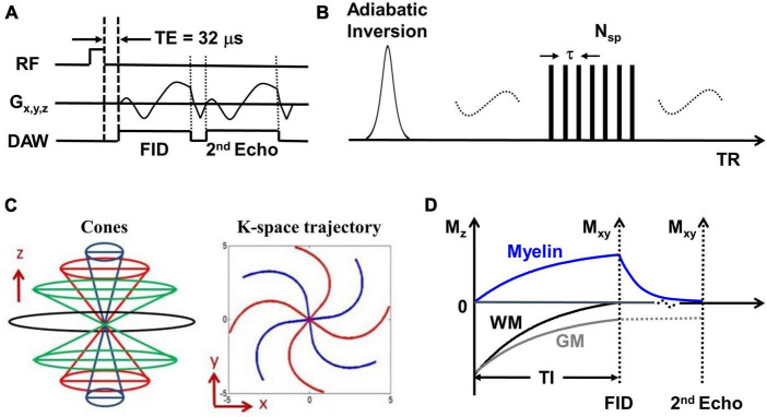
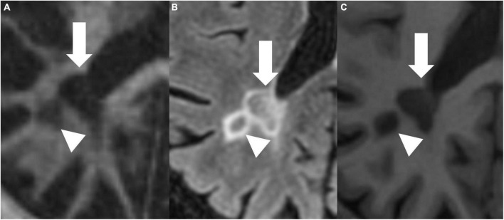
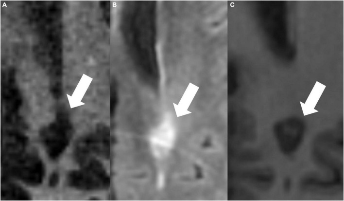
Twenty seven MS patients were included in this prospective study. IR-UTE, FLAIR, and MPRAGE sequences were employed on a 3T scanner. Regions of interest (ROIs) were manually drawn within the MS lesions, and the cerebrospinal fluid (CSF) and signal intensity ratios (SIR) were calculated from the derived values. Variations coefficients were determined from the standard deviations (Coeff 1) and the absolute differences (Coeff 2) of the SIRs. Disability grade was assessed by the expanded disability status scale (EDSS). Cortical/gray matter, subcortical, infratentorial, and spinal lesions were excluded.

RESULTS

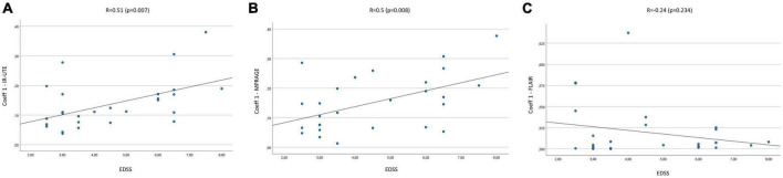
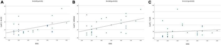
The mean diameter of the lesions was 7.8 ± 1.97 mm, while the mean EDSS score was 4.5 ± 1.73. We found moderate correlations between the EDSS and Coeff 1 and 2 on IR-UTE and MPRAGE images. Accordingly, Pearson's correlations on IR-UTE were = 0.51 ( = 0.007) and = 0.49 ( = 0.01) for Coeff 1 and 2, respectively. For MPRAGE, Pearson's correlations were = 0.5 ( = 0.008) and = 0.48 ( = 0.012) for Coeff 1 and 2, respectively. For FLAIR, only poor correlations could be found.

CONCLUSION

The SIVs of MS lesions on IR-UTE and MPRAGE images, assessed by Coeff 1 and 2, could be used as novel potential MRI biomarkers for patients' disability.

摘要

引言

尽管对多发性硬化症(MS)患者中许多基于病灶的MRI生物标志物进行了研究,但以往的研究均未涉及MS病灶的信号强度变化(SIV)。在本研究中,评估了直接髓鞘成像和标准临床序列上MS病灶的SIV,将其作为MS患者残疾程度的潜在MRI生物标志物。

方法

本前瞻性研究纳入了27例MS患者。在3T扫描仪上采用IR-UTE、FLAIR和MPRAGE序列。在MS病灶内手动绘制感兴趣区(ROI),并根据导出值计算脑脊液(CSF)和信号强度比(SIR)。变异系数由SIR的标准差(系数1)和绝对差值(系数2)确定。残疾程度采用扩展残疾状态量表(EDSS)进行评估。排除皮质/灰质、皮质下、幕下和脊髓病灶。

结果

病灶的平均直径为7.8±1.97mm,而平均EDSS评分为4.5±1.73。我们发现EDSS与IR-UTE和MPRAGE图像上的系数1和系数2之间存在中度相关性。因此,IR-UTE上系数1和系数2的Pearson相关性分别为r = 0.51(p = 0.007)和r = 0.49(p = 0.01)。对于MPRAGE,系数1和系数2的Pearson相关性分别为r = 0.5(p = 0.008)和r = 0.48(p = 0.012)。对于FLAIR,仅发现弱相关性。

结论

通过系数1和系数2评估的IR-UTE和MPRAGE图像上MS病灶的SIV,可作为评估患者残疾程度的新型潜在MRI生物标志物。

https://cdn.ncbi.nlm.nih.gov/pmc/blobs/ee60/10040653/3f9818858f2c/fnins-17-1145251-g005.jpg
https://cdn.ncbi.nlm.nih.gov/pmc/blobs/ee60/10040653/5a4394c20f34/fnins-17-1145251-g001.jpg
https://cdn.ncbi.nlm.nih.gov/pmc/blobs/ee60/10040653/a1f0d30fde63/fnins-17-1145251-g002.jpg
https://cdn.ncbi.nlm.nih.gov/pmc/blobs/ee60/10040653/69be45fbf142/fnins-17-1145251-g003.jpg
https://cdn.ncbi.nlm.nih.gov/pmc/blobs/ee60/10040653/8be22dbb5dfe/fnins-17-1145251-g004.jpg
https://cdn.ncbi.nlm.nih.gov/pmc/blobs/ee60/10040653/3f9818858f2c/fnins-17-1145251-g005.jpg

相似文献

1
The signal intensity variation of multiple sclerosis (MS) lesions on magnetic resonance imaging (MRI) as a potential biomarker for patients' disability: A feasibility study.磁共振成像(MRI)上多发性硬化症(MS)病灶的信号强度变化作为患者残疾的潜在生物标志物:一项可行性研究。
Front Neurosci. 2023 Mar 13;17:1145251. doi: 10.3389/fnins.2023.1145251. eCollection 2023.
2
Clinical evaluation of white matter lesions on 3D inversion recovery ultrashort echo time MRI in multiple sclerosis.三维反转恢复超短回波时间磁共振成像对多发性硬化症白质病变的临床评估
Quant Imaging Med Surg. 2023 Jul 1;13(7):4171-4180. doi: 10.21037/qims-22-1317. Epub 2023 May 4.
3
T1- Thresholds in Black Holes Increase Clinical-Radiological Correlation in Multiple Sclerosis Patients.T1 - 黑洞中的阈值增加了多发性硬化症患者的临床 - 放射学相关性。
PLoS One. 2015 Dec 11;10(12):e0144693. doi: 10.1371/journal.pone.0144693. eCollection 2015.
4
Detection and quantification of regional cortical gray matter damage in multiple sclerosis utilizing gradient echo MRI.利用梯度回波磁共振成像检测和量化多发性硬化症中局部皮质灰质损伤
Neuroimage Clin. 2015 Aug 18;9:164-75. doi: 10.1016/j.nicl.2015.08.003. eCollection 2015.
5
Glial and neuroaxonal biomarkers in a multiple sclerosis (MS) cohort.多发性硬化症(MS)队列中的神经胶质和神经轴突生物标志物
Hell J Nucl Med. 2019 Sep-Dec;22 Suppl 2:113-121.
6
Quantitative texture analysis of brain white matter lesions derived from T2-weighted MR images in MS patients with clinically isolated syndrome.对临床孤立综合征多发性硬化症患者T2加权磁共振图像中脑白质病变进行定量纹理分析。
J Neuroradiol. 2015 Apr;42(2):99-114. doi: 10.1016/j.neurad.2014.05.006. Epub 2014 Jun 23.
7
Association of volumetric MRI measures and disability in MS patients of the same age: Descriptions from a birth year cohort.磁共振容积成像测量与同龄 MS 患者残疾的相关性:来自同一年龄队列的描述。
Mult Scler Relat Disord. 2023 Mar;71:104568. doi: 10.1016/j.msard.2023.104568. Epub 2023 Feb 12.
8
Multiple sclerosis lesions and irreversible brain tissue damage: a comparative ultrahigh-field strength magnetic resonance imaging study.多发性硬化病变与不可逆性脑组织损伤:一项超高场强磁共振成像对比研究
Arch Neurol. 2012 Jun;69(6):739-45. doi: 10.1001/archneurol.2011.2450.
9
Extensive Brain Pathologic Alterations Detected with 7.0-T MR Spectroscopic Imaging Associated with Disability in Multiple Sclerosis.7.0T 磁共振波谱成像检测到的广泛脑病理改变与多发性硬化的残疾相关。
Radiology. 2022 Apr;303(1):141-150. doi: 10.1148/radiol.210614. Epub 2022 Jan 4.
10
[Magnetic resonance findings in the brain of patients with multiple sclerosis without oligoclonal bands in the cerebrospinal fluid].[脑脊液中无寡克隆带的多发性硬化症患者脑部的磁共振成像结果]
Srp Arh Celok Lek. 2003 Jan-Feb;131(1-2):31-5. doi: 10.2298/sarh0302031m.

引用本文的文献

1
Novel fat suppression technique for ultrashort echo time MRI using single-point Dixon phase modeling.使用单点狄克逊相位建模的超短回波时间磁共振成像新型脂肪抑制技术。
Quant Imaging Med Surg. 2025 May 1;15(5):4580-4591. doi: 10.21037/qims-24-1998. Epub 2025 Apr 10.
2
Hemosiderin quantification in hemophilic arthropathy using quantitative magnetic resonance imaging.利用定量磁共振成像对血友病性关节病中的含铁血黄素进行定量分析。
Sci Rep. 2025 Apr 24;15(1):14353. doi: 10.1038/s41598-025-99085-7.
3
An updated systematic review and meta-analysis exploring the efficacy and safety of dimethyl fumarate (DMF) for patients with multiple sclerosis (MS).

本文引用的文献

1
Direct imaging of white matter ultrashort T components at 7 Tesla.在 7 特斯拉下对白质超短 T 成分的直接成像。
Magn Reson Imaging. 2022 Feb;86:107-117. doi: 10.1016/j.mri.2021.11.016. Epub 2021 Dec 11.
2
Inversion Recovery Ultrashort TE MR Imaging of Myelin is Significantly Correlated with Disability in Patients with Multiple Sclerosis.反转恢复超短 TE 磁共振成像髓鞘与多发性硬化症患者的残疾显著相关。
AJNR Am J Neuroradiol. 2021 May;42(5):868-874. doi: 10.3174/ajnr.A7006. Epub 2021 Feb 18.
3
Volumetric imaging of myelin in vivo using 3D inversion recovery-prepared ultrashort echo time cones magnetic resonance imaging.
一项最新的系统评价和荟萃分析,探讨富马酸二甲酯(DMF)对多发性硬化症(MS)患者的疗效和安全性。
BMJ Neurol Open. 2024 Dec 18;6(2):e000872. doi: 10.1136/bmjno-2024-000872. eCollection 2024.
4
Predicting multiple sclerosis disease progression and outcomes with machine learning and MRI-based biomarkers: a review.利用机器学习和基于MRI的生物标志物预测多发性硬化症的疾病进展和预后:综述
J Neurol. 2024 Oct;271(10):6543-6572. doi: 10.1007/s00415-024-12651-3. Epub 2024 Sep 12.
5
Impact of dance classes on motor and cognitive functions and gut microbiota composition in multiple sclerosis patients: Randomized controlled trial.舞蹈课程对多发性硬化症患者运动和认知功能及肠道微生物群落组成的影响:随机对照试验。
Eur J Sport Sci. 2024 Aug;24(8):1186-1196. doi: 10.1002/ejsc.12166. Epub 2024 Jul 5.
6
Association of vulnerable plaques with white matter hyperintensities on high-resolution magnetic resonance imaging.高分辨率磁共振成像中易损斑块与脑白质高信号的关联。
Quant Imaging Med Surg. 2024 May 1;14(5):3606-3618. doi: 10.21037/qims-23-1856. Epub 2024 Apr 26.
7
Bibliometric analysis of myelin imaging studies of patients with multiple sclerosis (2000-2022).多发性硬化症患者髓鞘成像研究的文献计量分析(2000 - 2022年)
Quant Imaging Med Surg. 2024 Jan 3;14(1):837-851. doi: 10.21037/qims-23-1157. Epub 2024 Jan 2.
使用三维反转恢复准备的超短回波时间圆锥磁共振成像对体内髓磷脂进行容积成像。
NMR Biomed. 2020 Oct;33(10):e4326. doi: 10.1002/nbm.4326. Epub 2020 Jul 20.
4
So You Want to Image Myelin Using MRI: An Overview and Practical Guide for Myelin Water Imaging.那么你想用MRI对髓磷脂成像:髓磷脂水成像概述及实用指南。
J Magn Reson Imaging. 2021 Feb;53(2):360-373. doi: 10.1002/jmri.27059. Epub 2020 Feb 3.
5
Whole-Brain Myelin Imaging Using 3D Double-Echo Sliding Inversion Recovery Ultrashort Echo Time (DESIRE UTE) MRI.使用 3D 双回波滑动反转恢复超短回波时间(DESIRE UTE)MRI 进行全脑髓鞘成像。
Radiology. 2020 Feb;294(2):362-374. doi: 10.1148/radiol.2019190911. Epub 2019 Nov 19.
6
Association between pathological and MRI findings in multiple sclerosis.多发性硬化的病理与 MRI 表现的相关性。
Lancet Neurol. 2019 Feb;18(2):198-210. doi: 10.1016/S1474-4422(18)30451-4.
7
Multiple sclerosis: clinical aspects.多发性硬化症:临床方面。
Curr Opin Neurol. 2018 Dec;31(6):752-759. doi: 10.1097/WCO.0000000000000622.
8
Progressive multiple sclerosis patients show substantial lesion activity that correlates with clinical disease severity and sex: a retrospective autopsy cohort analysis.进展性多发性硬化症患者表现出大量的病变活动,与临床疾病严重程度和性别相关:一项回顾性尸检队列分析。
Acta Neuropathol. 2018 Apr;135(4):511-528. doi: 10.1007/s00401-018-1818-y. Epub 2018 Feb 13.
9
In vivo characterization of brain ultrashort-T components.脑超短 T 成分的体内特征描述。
Magn Reson Med. 2018 Aug;80(2):726-735. doi: 10.1002/mrm.27037. Epub 2017 Dec 1.
10
Short T imaging using a 3D double adiabatic inversion recovery prepared ultrashort echo time cones (3D DIR-UTE-Cones) sequence.使用三维双反转绝热恢复超短回波时间锥形(3D DIR-UTE-Cones)序列进行短 T 成像。
Magn Reson Med. 2018 May;79(5):2555-2563. doi: 10.1002/mrm.26908. Epub 2017 Sep 14.