• 文献检索
  • 文档翻译
  • 深度研究
  • 学术资讯
  • Suppr Zotero 插件Zotero 插件
  • 邀请有礼
  • 套餐&价格
  • 历史记录
应用&插件
Suppr Zotero 插件Zotero 插件浏览器插件Mac 客户端Windows 客户端微信小程序
定价
高级版会员购买积分包购买API积分包
服务
文献检索文档翻译深度研究API 文档MCP 服务
关于我们
关于 Suppr公司介绍联系我们用户协议隐私条款
关注我们

Suppr 超能文献

核心技术专利:CN118964589B侵权必究
粤ICP备2023148730 号-1Suppr @ 2026

文献检索

告别复杂PubMed语法,用中文像聊天一样搜索,搜遍4000万医学文献。AI智能推荐,让科研检索更轻松。

立即免费搜索

文件翻译

保留排版,准确专业,支持PDF/Word/PPT等文件格式,支持 12+语言互译。

免费翻译文档

深度研究

AI帮你快速写综述,25分钟生成高质量综述,智能提取关键信息,辅助科研写作。

立即免费体验

主动网格和神经网络流水线用于细胞聚集体分割。

Active mesh and neural network pipeline for cell aggregate segmentation.

机构信息

The Francis Crick Institute, London, United Kingdom.

Photonics Group, Department of Physics, Imperial College London, London, United Kingdom.

出版信息

Biophys J. 2023 May 2;122(9):1586-1599. doi: 10.1016/j.bpj.2023.03.038. Epub 2023 Mar 30.

DOI:10.1016/j.bpj.2023.03.038
PMID:37002604
原文链接:https://pmc.ncbi.nlm.nih.gov/articles/PMC10183373/
Abstract

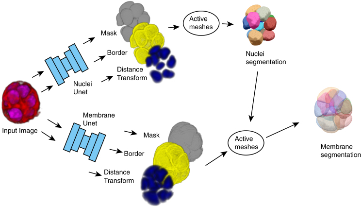
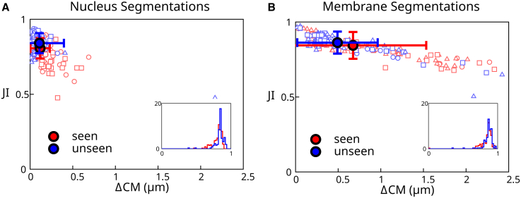
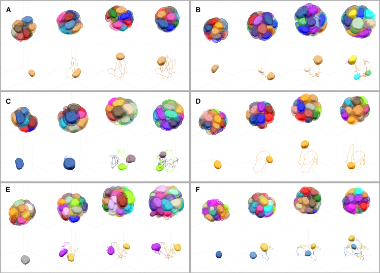
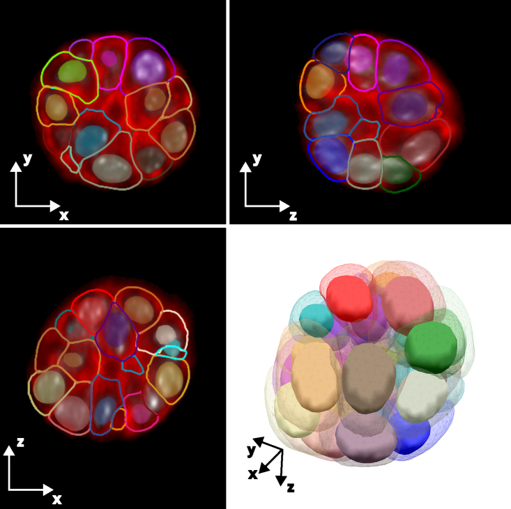
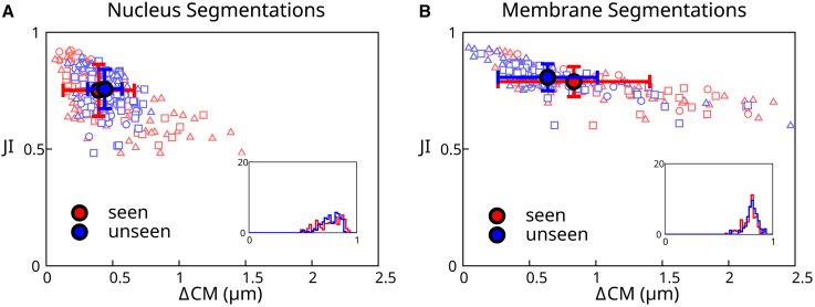
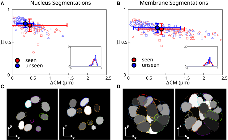
Segmenting cells within cellular aggregates in 3D is a growing challenge in cell biology due to improvements in capacity and accuracy of microscopy techniques. Here, we describe a pipeline to segment images of cell aggregates in 3D. The pipeline combines neural network segmentations with active meshes. We apply our segmentation method to cultured mouse mammary gland organoids imaged over 24 h with oblique plane microscopy, a high-throughput light-sheet fluorescence microscopy technique. We show that our method can also be applied to images of mouse embryonic stem cells imaged with a spinning disc microscope. We segment individual cells based on nuclei and cell membrane fluorescent markers, and track cells over time. We describe metrics to quantify the quality of the automated segmentation. Our segmentation pipeline involves a Fiji plugin that implements active mesh deformation and allows a user to create training data, automatically obtain segmentation meshes from original image data or neural network prediction, and manually curate segmentation data to identify and correct mistakes. Our active meshes-based approach facilitates segmentation postprocessing, correction, and integration with neural network prediction.

摘要

在细胞生物学中,由于显微镜技术的容量和准确性的提高,对细胞聚集体内的细胞进行分割是一个日益增长的挑战。在这里,我们描述了一个用于分割三维细胞聚集体图像的流水线。该流水线将神经网络分割与主动网格相结合。我们将我们的分割方法应用于经过 24 小时斜平面显微镜成像的培养的小鼠乳腺类器官,这是一种高通量光片荧光显微镜技术。我们表明,我们的方法也可以应用于使用旋转盘显微镜成像的小鼠胚胎干细胞的图像。我们基于细胞核和细胞膜荧光标记物对单个细胞进行分割,并随时间跟踪细胞。我们描述了用于量化自动分割质量的指标。我们的分割流水线涉及一个 Fiji 插件,该插件实现了主动网格变形,并允许用户创建训练数据,自动从原始图像数据或神经网络预测中获取分割网格,并手动编辑分割数据以识别和纠正错误。我们基于主动网格的方法便于分割后处理、校正以及与神经网络预测的集成。

https://cdn.ncbi.nlm.nih.gov/pmc/blobs/b795/10183373/0688fc45d078/gr10.jpg
https://cdn.ncbi.nlm.nih.gov/pmc/blobs/b795/10183373/91c3cfbf0f17/gr1.jpg
https://cdn.ncbi.nlm.nih.gov/pmc/blobs/b795/10183373/3bfa9a98d4f3/gr2.jpg
https://cdn.ncbi.nlm.nih.gov/pmc/blobs/b795/10183373/8c35470ac4f2/gr3.jpg
https://cdn.ncbi.nlm.nih.gov/pmc/blobs/b795/10183373/6b254f712598/gr4.jpg
https://cdn.ncbi.nlm.nih.gov/pmc/blobs/b795/10183373/e562fb8b891b/gr5.jpg
https://cdn.ncbi.nlm.nih.gov/pmc/blobs/b795/10183373/b121040f5cff/gr6.jpg
https://cdn.ncbi.nlm.nih.gov/pmc/blobs/b795/10183373/adad2acb5713/gr7.jpg
https://cdn.ncbi.nlm.nih.gov/pmc/blobs/b795/10183373/7e9f7c4e9296/gr8.jpg
https://cdn.ncbi.nlm.nih.gov/pmc/blobs/b795/10183373/c413e5b30a67/gr9.jpg
https://cdn.ncbi.nlm.nih.gov/pmc/blobs/b795/10183373/0688fc45d078/gr10.jpg

相似文献

1
Active mesh and neural network pipeline for cell aggregate segmentation.主动网格和神经网络流水线用于细胞聚集体分割。
Biophys J. 2023 May 2;122(9):1586-1599. doi: 10.1016/j.bpj.2023.03.038. Epub 2023 Mar 30.
2
Application of convolutional neural networks towards nuclei segmentation in localization-based super-resolution fluorescence microscopy images.基于定位的超分辨率荧光显微镜图像中核分割的卷积神经网络应用。
BMC Bioinformatics. 2021 Jun 15;22(1):325. doi: 10.1186/s12859-021-04245-x.
3
Bi-channel image registration and deep-learning segmentation (BIRDS) for efficient, versatile 3D mapping of mouse brain.双通道图像配准和深度学习分割(BIRDS)用于高效、通用的小鼠大脑 3D 映射。
Elife. 2021 Jan 18;10:e63455. doi: 10.7554/eLife.63455.
4
Automatic three-dimensional segmentation of mouse embryonic stem cell nuclei by utilising multiple channels of confocal fluorescence images.利用共聚焦荧光图像的多个通道自动对小鼠胚胎干细胞核进行三维分割。
J Microsc. 2021 Jan;281(1):57-75. doi: 10.1111/jmi.12949. Epub 2020 Aug 8.
5
Brain tumor segmentation and detection in MRI using convolutional neural networks and VGG16.使用卷积神经网络和VGG16在磁共振成像(MRI)中进行脑肿瘤分割与检测
Cancer Biomark. 2025 Mar;42(3):18758592241311184. doi: 10.1177/18758592241311184. Epub 2025 Apr 4.
6
Mandible segmentation from CT data for virtual surgical planning using an augmented two-stepped convolutional neural network.使用增强型两步卷积神经网络对 CT 数据进行下颌骨分割,用于虚拟手术规划。
Int J Comput Assist Radiol Surg. 2023 Aug;18(8):1479-1488. doi: 10.1007/s11548-022-02830-w. Epub 2023 Jan 13.
7
SUSAN: segment unannotated image structure using adversarial network.苏珊:使用对抗网络分割未注释的图像结构。
Magn Reson Med. 2019 May;81(5):3330-3345. doi: 10.1002/mrm.27627. Epub 2018 Dec 10.
8
A robust algorithm for segmenting and tracking clustered cells in time-lapse fluorescent microscopy.一种用于在荧光显微镜延时拍摄中分割和跟踪聚集细胞的稳健算法。
IEEE J Biomed Health Inform. 2013 Jul;17(4):862-9. doi: 10.1109/JBHI.2013.2262233.
9
Empirical gradient threshold technique for automated segmentation across image modalities and cell lines.用于跨图像模态和细胞系自动分割的经验梯度阈值技术。
J Microsc. 2015 Oct;260(1):86-99. doi: 10.1111/jmi.12269. Epub 2015 Jun 5.
10
Micro-Net: A unified model for segmentation of various objects in microscopy images.微网:用于显微镜图像中各种对象分割的统一模型。
Med Image Anal. 2019 Feb;52:160-173. doi: 10.1016/j.media.2018.12.003. Epub 2018 Dec 15.

引用本文的文献

1
Using the TSA-LSTM two-stage model to predict cancer incidence and mortality.使用TSA-LSTM两阶段模型预测癌症发病率和死亡率。
PLoS One. 2025 Feb 20;20(2):e0317148. doi: 10.1371/journal.pone.0317148. eCollection 2025.
2
Automated 3D cytoplasm segmentation in soft X-ray tomography.软X射线断层扫描中的自动化3D细胞质分割
iScience. 2024 Apr 29;27(6):109856. doi: 10.1016/j.isci.2024.109856. eCollection 2024 Jun 21.
3
Universal consensus 3D segmentation of cells from 2D segmented stacks.从二维分割堆栈中对细胞进行通用一致的三维分割。

本文引用的文献

1
Multiscale light-sheet organoid imaging framework.多尺度光片器官成像框架。
Nat Commun. 2022 Aug 18;13(1):4864. doi: 10.1038/s41467-022-32465-z.
2
EmbedSeg: Embedding-based Instance Segmentation for Biomedical Microscopy Data.EmbedSeg:基于嵌入的生物医学显微镜数据实例分割。
Med Image Anal. 2022 Oct;81:102523. doi: 10.1016/j.media.2022.102523. Epub 2022 Jul 3.
3
Mechanical constraints to cell-cycle progression in a pseudostratified epithelium.细胞周期在假复层上皮中的机械限制。
bioRxiv. 2025 Mar 20:2024.05.03.592249. doi: 10.1101/2024.05.03.592249.
4
Probing organoid metabolism using fluorescence lifetime imaging microscopy (FLIM): The next frontier of drug discovery and disease understanding.利用荧光寿命成像显微镜(FLIM)探测类器官代谢:药物发现和疾病认识的下一个前沿。
Adv Drug Deliv Rev. 2023 Oct;201:115081. doi: 10.1016/j.addr.2023.115081. Epub 2023 Aug 28.
Curr Biol. 2022 May 9;32(9):2076-2083.e2. doi: 10.1016/j.cub.2022.03.004. Epub 2022 Mar 25.
4
Tracking cell lineages in 3D by incremental deep learning.通过渐进式深度学习追踪 3D 中的细胞谱系。
Elife. 2022 Jan 6;11:e69380. doi: 10.7554/eLife.69380.
5
Dual-view oblique plane microscopy (dOPM).双视图斜平面显微镜检查(dOPM)。
Biomed Opt Express. 2020 Nov 18;11(12):7204-7220. doi: 10.1364/BOE.409781. eCollection 2020 Dec 1.
6
Cellpose: a generalist algorithm for cellular segmentation.Cellpose:一种通用的细胞分割算法。
Nat Methods. 2021 Jan;18(1):100-106. doi: 10.1038/s41592-020-01018-x. Epub 2020 Dec 14.
7
Establishment of a morphological atlas of the Caenorhabditis elegans embryo using deep-learning-based 4D segmentation.利用基于深度学习的 4D 分割技术建立秀丽隐杆线虫胚胎形态图谱。
Nat Commun. 2020 Dec 7;11(1):6254. doi: 10.1038/s41467-020-19863-x.
8
OrganoidTracker: Efficient cell tracking using machine learning and manual error correction.类器官追踪器:使用机器学习和手动纠错进行高效的细胞追踪。
PLoS One. 2020 Oct 22;15(10):e0240802. doi: 10.1371/journal.pone.0240802. eCollection 2020.
9
Accurate and versatile 3D segmentation of plant tissues at cellular resolution.以细胞分辨率准确且灵活地对植物组织进行三维分割。
Elife. 2020 Jul 29;9:e57613. doi: 10.7554/eLife.57613.
10
Contact area-dependent cell communication and the morphological invariance of ascidian embryogenesis.接触面积依赖性细胞通讯与海鞘胚胎发生的形态不变性。
Science. 2020 Jul 10;369(6500). doi: 10.1126/science.aar5663.