Department of Orthodontics, The Affiliated Stomatological Hospital of Nanjing Medical University, Nanjing, 210029, China.
Jiangsu Province Key Laboratory of Oral Diseases, Nanjing Medical University, Nanjing, 210029, China.
BMC Oral Health. 2023 Apr 1;23(1):191. doi: 10.1186/s12903-023-02881-8.
The purpose of this study was to evaluate the accuracy of automatic cephalometric landmark localization and measurements using cephalometric analysis via artificial intelligence (AI) compared with computer-assisted manual analysis.
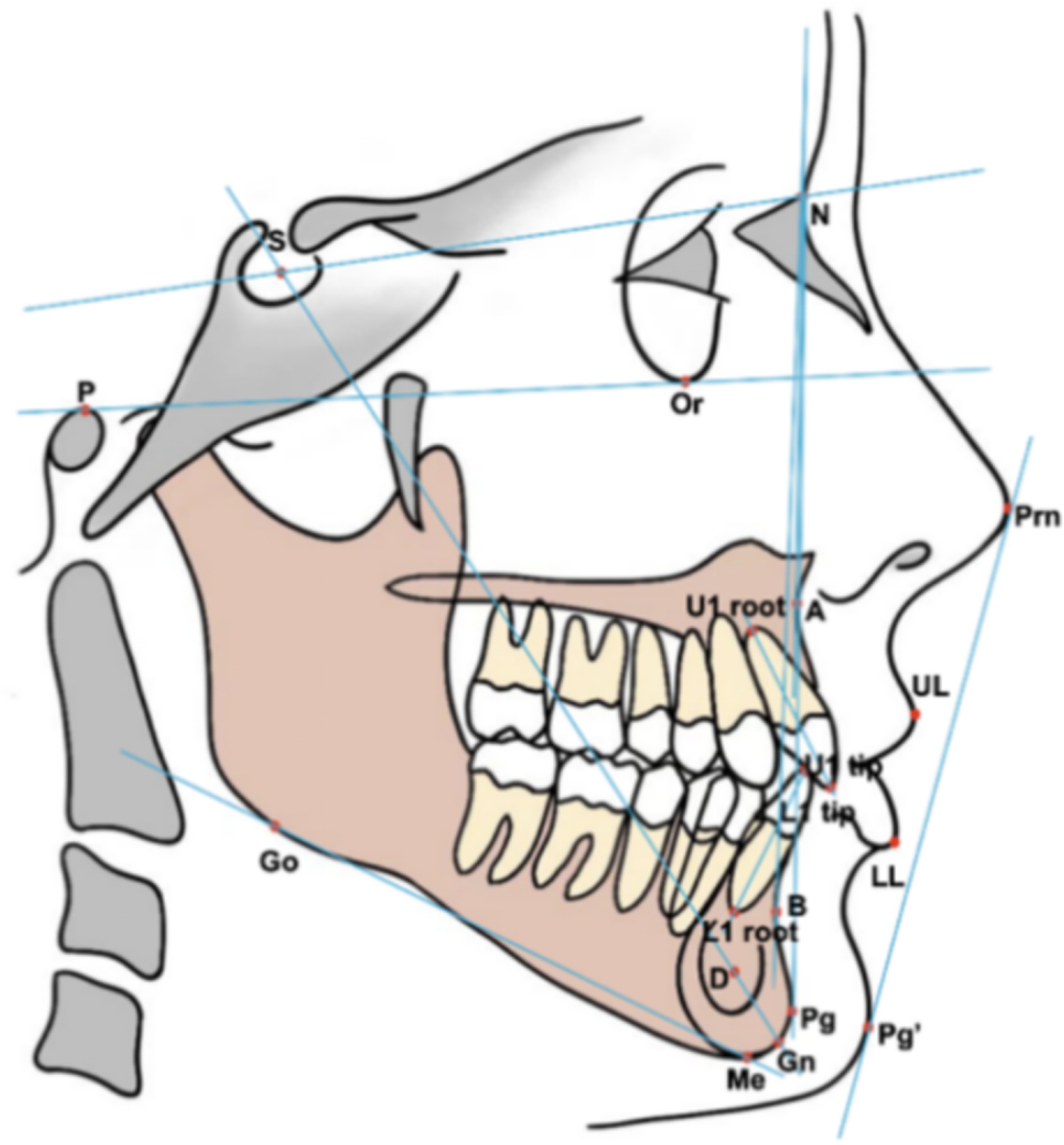
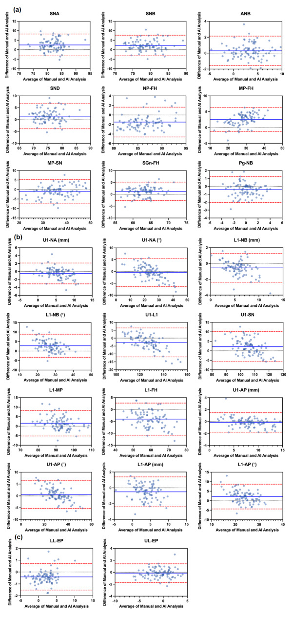
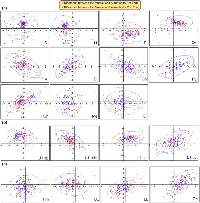
Reconstructed lateral cephalograms (RLCs) from cone-beam computed tomography (CBCT) in 85 patients were selected. Computer-assisted manual analysis (Dolphin Imaging 11.9) and AI automatic analysis (Planmeca Romexis 6.2) were used to locate 19 landmarks and obtain 23 measurements. Mean radial error (MRE) and successful detection rate (SDR) values were calculated to assess the accuracy of automatic landmark digitization. Paired t tests and Bland‒Altman plots were used to compare the differences and consistencies in cephalometric measurements between manual and automatic analysis programs.
The MRE for 19 cephalometric landmarks was 2.07 ± 1.35 mm with the automatic program. The average SDR within 1 mm, 2 mm, 2.5 mm, 3 and 4 mm were 18.82%, 58.58%, 71.70%, 82.04% and 91.39%, respectively. Soft tissue landmarks (1.54 ± 0.85 mm) had the most consistency, while dental landmarks (2.37 ± 1.55 mm) had the most variation. In total, 15 out of 23 measurements were within the clinically acceptable level of accuracy, 2 mm or 2°. The rates of consistency within the 95% limits of agreement were all above 90% for all measurement parameters.
Automatic analysis software collects cephalometric measurements almost effectively enough to be acceptable in clinical work. Nevertheless, automatic cephalometry is not capable of completely replacing manual tracing. Additional manual supervision and adjustment for automatic programs can increase accuracy and efficiency.
本研究旨在评估基于人工智能的头影测量分析(cephalometric analysis via artificial intelligence,CAI)自动定位和测量头影测量标志点的准确性,并与计算机辅助手动分析(computer-assisted manual analysis,CAM)进行比较。
选择 85 例患者的锥形束 CT(cone-beam computed tomography,CBCT)重建的侧位头颅定位片(reconstructed lateral cephalograms,RLCs)。使用计算机辅助手动分析软件(Dolphin Imaging 11.9)和 AI 自动分析软件(Planmeca Romexis 6.2)分别定位 19 个标志点并获取 23 个测量值。计算平均辐射误差(mean radial error,MRE)和成功检测率(success detection rate,SDR)以评估自动标志点数字化的准确性。采用配对 t 检验和 Bland–Altman 图比较手动和自动分析程序之间头影测量值的差异和一致性。
自动分析软件定位 19 个头影测量标志点的平均 MRE 为 2.07 ± 1.35 mm。平均 SDR 在 1、2、2.5、3 和 4 mm 范围内分别为 18.82%、58.58%、71.70%、82.04%和 91.39%。软组织标志点(1.54 ± 0.85 mm)的一致性最高,而牙标志点(2.37 ± 1.55 mm)的变化最大。总共 23 个测量值中有 15 个在临床可接受的精度范围内,即 2 毫米或 2°。所有测量参数的 95%一致性界限内的一致性率均在 90%以上。
自动分析软件获取的头影测量值在临床工作中几乎可以接受,足够有效。然而,自动头影测量术不能完全取代手动描记。对自动程序进行额外的手动监督和调整可以提高准确性和效率。