Gao Jianxiong, Niu Rong, Shi Yunmei, Shao Xiaoliang, Jiang Zhenxing, Ge Xinyu, Wang Yuetao, Shao Xiaonan
Department of Nuclear Medicine, The Third Affiliated Hospital of Soochow University, Changzhou, 213003, China.
Institute of Clinical Translation of Nuclear Medicine and Molecular Imaging, Soochow University, Changzhou, 213003, China.
EJNMMI Res. 2023 Apr 4;13(1):26. doi: 10.1186/s13550-023-00977-4.
This study aims to construct radiomics models based on [F]FDG PET/CT using multiple machine learning methods to predict the EGFR mutation status of lung adenocarcinoma and evaluate whether incorporating clinical parameters can improve the performance of radiomics models.
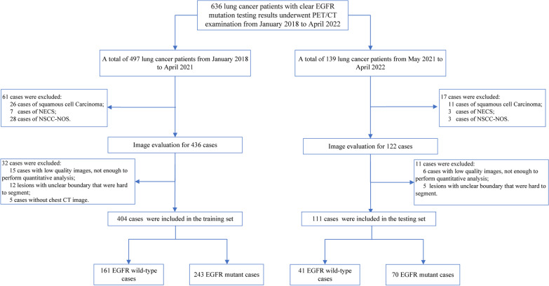
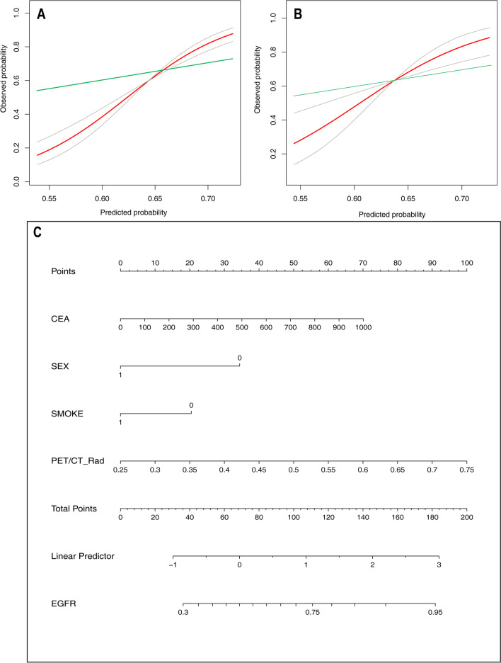
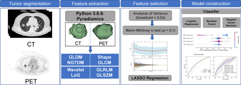
A total of 515 patients were retrospectively collected and divided into a training set (n = 404) and an independent testing set (n = 111) according to their examination time. After semi-automatic segmentation of PET/CT images, the radiomics features were extracted, and the best feature sets of CT, PET, and PET/CT modalities were screened out. Nine radiomics models were constructed using logistic regression (LR), random forest (RF), and support vector machine (SVM) methods. According to the performance in the testing set, the best model of the three modalities was kept, and its radiomics score (Rad-score) was calculated. Furthermore, combined with the valuable clinical parameters (gender, smoking history, nodule type, CEA, SCC-Ag), a joint radiomics model was built.
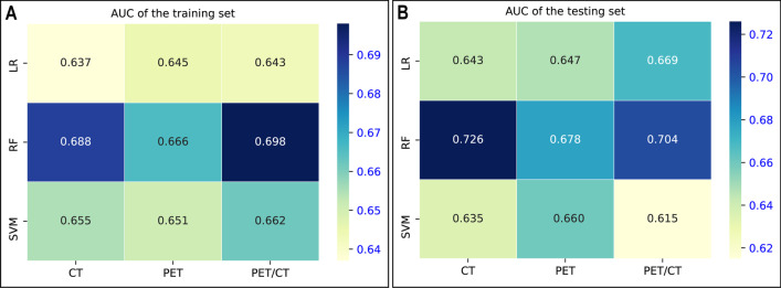
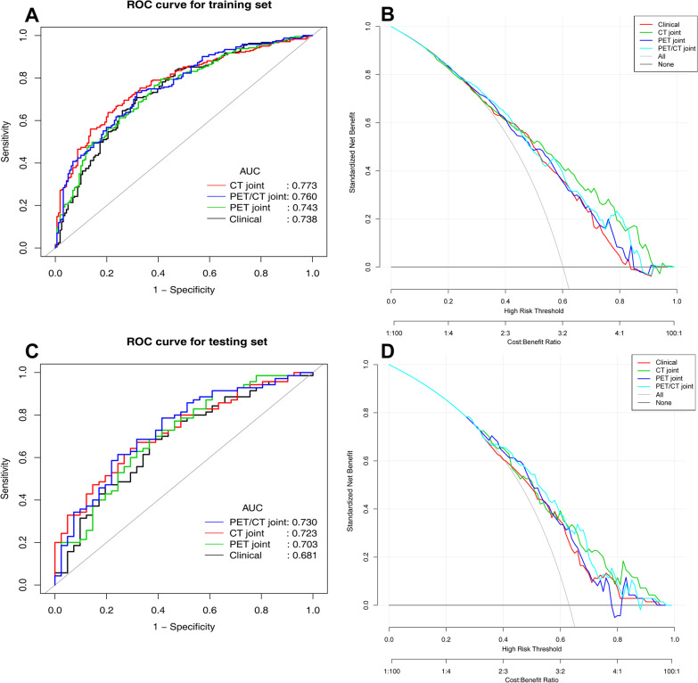
Compared with LR and SVM, the RF Rad-score showed the best performance among the three radiomics models of CT, PET, and PET/CT (training and testing sets AUC: 0.688, 0.666, and 0.698 vs. 0.726, 0.678, and 0.704). Among the three joint models, the PET/CT joint model performed the best (training and testing sets AUC: 0.760 vs. 0.730). The further stratified analysis found that CT_RF had the best prediction effect for stage I-II lesions (training set and testing set AUC: 0.791 vs. 0.797), while PET/CT joint model had the best prediction effect for stage III-IV lesions (training and testing sets AUC: 0.722 vs. 0.723).
Combining with clinical parameters can improve the predictive performance of PET/CT radiomics model, especially for patients with advanced lung adenocarcinoma.
本研究旨在基于[F]FDG PET/CT,使用多种机器学习方法构建放射组学模型,以预测肺腺癌的表皮生长因子受体(EGFR)突变状态,并评估纳入临床参数是否能提高放射组学模型的性能。
回顾性收集515例患者,根据检查时间将其分为训练集(n = 404)和独立测试集(n = 111)。对PET/CT图像进行半自动分割后,提取放射组学特征,并筛选出CT、PET和PET/CT模态的最佳特征集。使用逻辑回归(LR)、随机森林(RF)和支持向量机(SVM)方法构建9个放射组学模型。根据测试集中的表现,保留三种模态中最佳的模型,并计算其放射组学评分(Rad-score)。此外,结合有价值的临床参数(性别、吸烟史、结节类型、癌胚抗原、鳞状细胞癌抗原),构建联合放射组学模型。
与LR和SVM相比,RF Rad-score在CT、PET和PET/CT的三种放射组学模型中表现最佳(训练集和测试集的曲线下面积[AUC]:分别为0.688、0.666、0.698和0.726、0.678、0.704)。在三种联合模型中,PET/CT联合模型表现最佳(训练集和测试集AUC:分别为0.760和0.730)。进一步的分层分析发现,CT_RF对I-II期病变的预测效果最佳(训练集和测试集AUC:分别为0.791和0.797),而PET/CT联合模型对III-IV期病变的预测效果最佳(训练集和测试集AUC:分别为0.722和0.723)。
结合临床参数可提高PET/CT放射组学模型的预测性能,尤其是对于晚期肺腺癌患者。