• 文献检索
  • 文档翻译
  • 深度研究
  • 学术资讯
  • Suppr Zotero 插件Zotero 插件
  • 邀请有礼
  • 套餐&价格
  • 历史记录
应用&插件
Suppr Zotero 插件Zotero 插件浏览器插件Mac 客户端Windows 客户端微信小程序
定价
高级版会员购买积分包购买API积分包
服务
文献检索文档翻译深度研究API 文档MCP 服务
关于我们
关于 Suppr公司介绍联系我们用户协议隐私条款
关注我们

Suppr 超能文献

核心技术专利:CN118964589B侵权必究
粤ICP备2023148730 号-1Suppr @ 2026

文献检索

告别复杂PubMed语法,用中文像聊天一样搜索,搜遍4000万医学文献。AI智能推荐,让科研检索更轻松。

立即免费搜索

文件翻译

保留排版,准确专业,支持PDF/Word/PPT等文件格式,支持 12+语言互译。

免费翻译文档

深度研究

AI帮你快速写综述,25分钟生成高质量综述,智能提取关键信息,辅助科研写作。

立即免费体验

用于关联体积电子显微镜的基因编码条码。

Genetically encoded barcodes for correlative volume electron microscopy.

机构信息

Munich Institute of Biomedical Engineering, Department of Bioscience, TUM School of Natural Sciences and TUM School of Medicine, Technical University of Munich, Munich, Germany.

Institute for Synthetic Biomedicine, Helmholtz Munich, Neuherberg, Germany.

出版信息

Nat Biotechnol. 2023 Dec;41(12):1734-1745. doi: 10.1038/s41587-023-01713-y. Epub 2023 Apr 17.

DOI:10.1038/s41587-023-01713-y
PMID:37069313
原文链接:https://pmc.ncbi.nlm.nih.gov/articles/PMC10713455/
Abstract

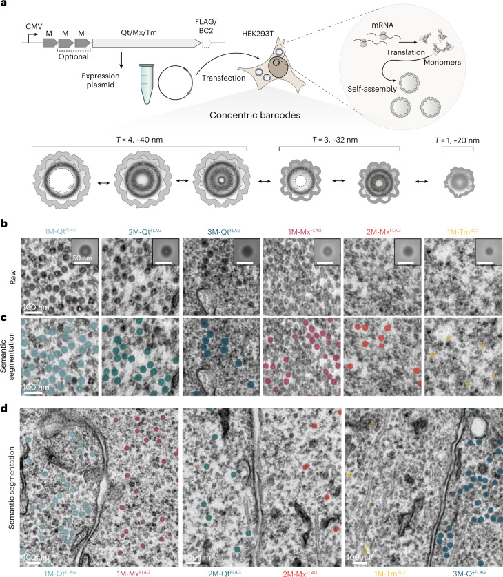
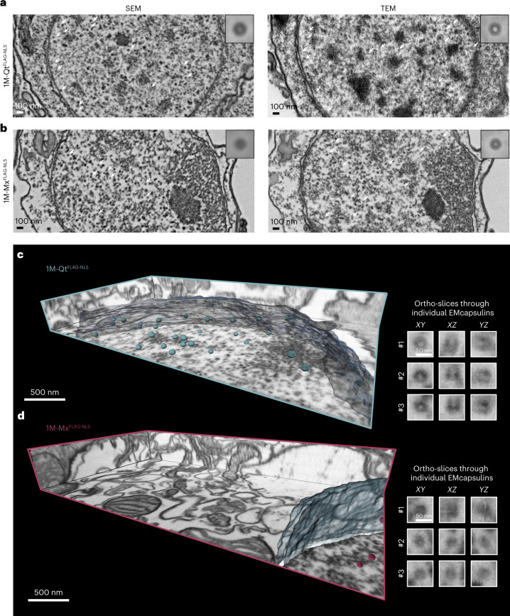
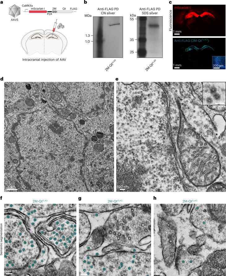
While genetically encoded reporters are common for fluorescence microscopy, equivalent multiplexable gene reporters for electron microscopy (EM) are still scarce. Here, by installing a variable number of fixation-stable metal-interacting moieties in the lumen of encapsulin nanocompartments of different sizes, we developed a suite of spherically symmetric and concentric barcodes (EMcapsulins) that are readable by standard EM techniques. Six classes of EMcapsulins could be automatically segmented and differentiated. The coding capacity was further increased by arranging several EMcapsulins into distinct patterns via a set of rigid spacers of variable length. Fluorescent EMcapsulins were expressed to monitor subcellular structures in light and EM. Neuronal expression in Drosophila and mouse brains enabled the automatic identification of genetically defined cells in EM. EMcapsulins are compatible with transmission EM, scanning EM and focused ion beam scanning EM. The expandable palette of genetically controlled EM-readable barcodes can augment anatomical EM images with multiplexed gene expression maps.

摘要

虽然遗传编码报告基因常用于荧光显微镜,但适用于电子显微镜(EM)的可多重检测的基因报告基因仍然稀缺。在这里,我们通过在不同大小的 encapsulin 纳米隔室的腔室内安装数量可变的固定稳定的金属相互作用部分,开发了一系列具有球形对称和同心条带的条码(EMcapsulins),可通过标准 EM 技术进行读取。可以自动对六类 EMcapsulins 进行分割和区分。通过使用一组具有可变长度的刚性间隔物将几个 EMcapsulins 排列成不同的模式,进一步增加了编码容量。荧光 EMcapsulins 的表达可用于在光镜和 EM 下监测亚细胞结构。在果蝇和小鼠大脑中的神经元表达使我们能够在 EM 中自动识别遗传定义的细胞。EMcapsulins 与透射 EM、扫描 EM 和聚焦离子束扫描 EM 兼容。可扩展的遗传控制的 EM 可读条码调色板可以为解剖学 EM 图像添加多重基因表达图谱。

https://cdn.ncbi.nlm.nih.gov/pmc/blobs/02f6/10713455/781f51c34a1a/41587_2023_1713_Fig13_ESM.jpg
https://cdn.ncbi.nlm.nih.gov/pmc/blobs/02f6/10713455/ee4fe2fb03fd/41587_2023_1713_Fig1_HTML.jpg
https://cdn.ncbi.nlm.nih.gov/pmc/blobs/02f6/10713455/f347ae3abc98/41587_2023_1713_Fig2_HTML.jpg
https://cdn.ncbi.nlm.nih.gov/pmc/blobs/02f6/10713455/02409396aba7/41587_2023_1713_Fig3_HTML.jpg
https://cdn.ncbi.nlm.nih.gov/pmc/blobs/02f6/10713455/7f9637514704/41587_2023_1713_Fig4_HTML.jpg
https://cdn.ncbi.nlm.nih.gov/pmc/blobs/02f6/10713455/884c9f3ea89d/41587_2023_1713_Fig5_HTML.jpg
https://cdn.ncbi.nlm.nih.gov/pmc/blobs/02f6/10713455/d010d83278e7/41587_2023_1713_Fig6_HTML.jpg
https://cdn.ncbi.nlm.nih.gov/pmc/blobs/02f6/10713455/cfa6802f7414/41587_2023_1713_Fig7_ESM.jpg
https://cdn.ncbi.nlm.nih.gov/pmc/blobs/02f6/10713455/092faa7c682b/41587_2023_1713_Fig8_ESM.jpg
https://cdn.ncbi.nlm.nih.gov/pmc/blobs/02f6/10713455/d3b647be2147/41587_2023_1713_Fig9_ESM.jpg
https://cdn.ncbi.nlm.nih.gov/pmc/blobs/02f6/10713455/215bd5af6ec7/41587_2023_1713_Fig10_ESM.jpg
https://cdn.ncbi.nlm.nih.gov/pmc/blobs/02f6/10713455/c504116da154/41587_2023_1713_Fig11_ESM.jpg
https://cdn.ncbi.nlm.nih.gov/pmc/blobs/02f6/10713455/c2129163e8fc/41587_2023_1713_Fig12_ESM.jpg
https://cdn.ncbi.nlm.nih.gov/pmc/blobs/02f6/10713455/781f51c34a1a/41587_2023_1713_Fig13_ESM.jpg

相似文献

1
Genetically encoded barcodes for correlative volume electron microscopy.用于关联体积电子显微镜的基因编码条码。
Nat Biotechnol. 2023 Dec;41(12):1734-1745. doi: 10.1038/s41587-023-01713-y. Epub 2023 Apr 17.
2
Volume-CLEM: a method for correlative light and electron microscopy in three dimensions.Volume-CLEM:一种用于三维共聚焦和电子显微镜相关的方法。
Am J Physiol Lung Cell Mol Physiol. 2019 Dec 1;317(6):L778-L784. doi: 10.1152/ajplung.00333.2019. Epub 2019 Sep 11.
3
Genetically targeted 3D visualisation of Drosophila neurons under Electron Microscopy and X-Ray Microscopy using miniSOG.利用 miniSOG 在电子显微镜和 X 射线显微镜下对果蝇神经元进行基因靶向 3D 可视化。
Sci Rep. 2016 Dec 13;6:38863. doi: 10.1038/srep38863.
4
VIPER is a genetically encoded peptide tag for fluorescence and electron microscopy.VIPER 是一种用于荧光和电子显微镜的基因编码肽标签。
Proc Natl Acad Sci U S A. 2018 Dec 18;115(51):12961-12966. doi: 10.1073/pnas.1808626115. Epub 2018 Dec 5.
5
FIB-SEM imaging properties of Drosophila melanogaster tissues embedded in Lowicryl HM20.在低聚甲醛 HM20 中包埋的果蝇组织的 FIB-SEM 成像特性。
J Microsc. 2019 Feb;273(2):91-104. doi: 10.1111/jmi.12764. Epub 2018 Nov 12.
6
Reconstructing neural circuits using multiresolution correlated light and electron microscopy.使用多分辨率相关光和电子显微镜重建神经回路。
Front Neural Circuits. 2022 Oct 21;16:753496. doi: 10.3389/fncir.2022.753496. eCollection 2022.
7
Correlative light and volume electron microscopy to study brain development.相关光镜和电镜技术研究脑发育。
Microscopy (Oxf). 2023 Aug 4;72(4):279-286. doi: 10.1093/jmicro/dfad002.
8
Modern field emission scanning electron microscopy provides new perspectives for imaging kidney ultrastructure.现代场发射扫描电子显微镜为观察肾脏超微结构提供了新的视角。
Kidney Int. 2018 Sep;94(3):625-631. doi: 10.1016/j.kint.2018.05.017.
9
Large-scale Scanning Transmission Electron Microscopy (Nanotomy) of Healthy and Injured Zebrafish Brain.健康和受伤斑马鱼大脑的大规模扫描透射电子显微镜(纳米断层成像)
J Vis Exp. 2016 May 25(111):53635. doi: 10.3791/53635.
10
Dual-labeling method for electron microscopy to characterize synaptic connectivity using genetically encoded fluorescent reporters in Drosophila.双标记方法用于电子显微镜,使用果蝇中遗传编码的荧光报告基因来描述突触连接。
J Neurosci Methods. 2011 Jan 15;194(2):312-5. doi: 10.1016/j.jneumeth.2010.10.020. Epub 2010 Nov 11.

引用本文的文献

1
Systematic Comparison of Commercial Uranyl-Alternative Stains for Negative- and Positive-Staining Transmission Electron Microscopy of Organic Specimens.用于有机标本负染色和正染色透射电子显微镜的商业铀替代染色剂的系统比较
Adv Healthc Mater. 2025 Jun;14(16):e2404870. doi: 10.1002/adhm.202404870. Epub 2025 Apr 29.
2
Cryo-electron tomography pipeline for plasma membranes.用于质膜的冷冻电子断层扫描流程
Nat Commun. 2025 Jan 20;16(1):855. doi: 10.1038/s41467-025-56045-z.
3
Cryo-electron tomography pipeline for plasma membranes.用于质膜的冷冻电子断层扫描流程

本文引用的文献

1
Spatial proteomics in three-dimensional intact specimens.三维完整标本中的空间蛋白质组学。
Cell. 2022 Dec 22;185(26):5040-5058.e19. doi: 10.1016/j.cell.2022.11.021.
2
Development and external validation of an MRI-based neural network for brain metastasis segmentation in the AURORA multicenter study.AURORA多中心研究中基于MRI的神经网络用于脑转移瘤分割的开发与外部验证
Radiother Oncol. 2023 Jan;178:109425. doi: 10.1016/j.radonc.2022.11.014. Epub 2022 Nov 26.
3
An open-access volume electron microscopy atlas of whole cells and tissues.
bioRxiv. 2024 Jun 28:2024.06.27.600657. doi: 10.1101/2024.06.27.600657.
4
Multiplexed volumetric CLEM enabled by scFvs provides insights into the cytology of cerebellar cortex.基于 scFv 的多重体积 CLEM 使小脑皮质细胞学研究成为可能。
Nat Commun. 2024 Aug 5;15(1):6648. doi: 10.1038/s41467-024-50411-z.
5
Bioorthogonal Labeling Enables In Situ Fluorescence Imaging of Expressed Gas Vesicle Nanostructures.生物正交标记可实现表达气穴纳米结构的原位荧光成像。
Bioconjug Chem. 2024 Mar 20;35(3):333-339. doi: 10.1021/acs.bioconjchem.3c00518. Epub 2024 Feb 12.
6
Genetically encoded multimeric tags for subcellular protein localization in cryo-EM.用于冷冻电镜中亚细胞蛋白定位的基因编码多聚标签。
Nat Methods. 2023 Dec;20(12):1900-1908. doi: 10.1038/s41592-023-02053-0. Epub 2023 Nov 6.
7
Magnetic and Fluorescent Dual-Labeled Genetically Encoded Targeted Nanoparticles for Malignant Glioma Cell Tracking and Drug Delivery.用于恶性胶质瘤细胞追踪与药物递送的磁性和荧光双标记基因编码靶向纳米颗粒
Pharmaceutics. 2023 Oct 4;15(10):2422. doi: 10.3390/pharmaceutics15102422.
开放获取的全细胞和组织电子显微镜图谱集
Nature. 2021 Nov;599(7883):147-151. doi: 10.1038/s41586-021-03992-4. Epub 2021 Oct 6.
4
Whole-cell organelle segmentation in volume electron microscopy.体积电子显微镜中的全细胞细胞器分割
Nature. 2021 Nov;599(7883):141-146. doi: 10.1038/s41586-021-03977-3. Epub 2021 Oct 6.
5
Non-invasive and high-throughput interrogation of exon-specific isoform expression.非侵入性和高通量的外显子特异性异构体表达检测。
Nat Cell Biol. 2021 Jun;23(6):652-663. doi: 10.1038/s41556-021-00678-x. Epub 2021 Jun 3.
6
Fragment-linking peptide design yields a high-affinity ligand for microtubule-based transport.片段连接肽设计产生一种基于微管的运输的高亲和力配体。
Cell Chem Biol. 2021 Sep 16;28(9):1347-1355.e5. doi: 10.1016/j.chembiol.2021.03.010. Epub 2021 Apr 9.
7
CEM500K, a large-scale heterogeneous unlabeled cellular electron microscopy image dataset for deep learning.CEM500K,一个用于深度学习的大规模异质无标签细胞电子显微镜图像数据集。
Elife. 2021 Apr 8;10:e65894. doi: 10.7554/eLife.65894.
8
nnU-Net: a self-configuring method for deep learning-based biomedical image segmentation.nnU-Net:一种基于深度学习的生物医学图像分割的自配置方法。
Nat Methods. 2021 Feb;18(2):203-211. doi: 10.1038/s41592-020-01008-z. Epub 2020 Dec 7.
9
Genetically encoded tags for direct synthesis of EM-visible gold nanoparticles in cells.用于在细胞中直接合成 EM 可见金纳米粒子的基因编码标签。
Nat Methods. 2020 Sep;17(9):937-946. doi: 10.1038/s41592-020-0911-z. Epub 2020 Aug 10.
10
LoTToR: An Algorithm for Missing-Wedge Correction of the Low-Tilt Tomographic 3D Reconstruction of a Single-Molecule Structure.LoTToR:一种用于单分子结构低倾度断层扫描 3D 重建的缺角校正算法。
Sci Rep. 2020 Jun 26;10(1):10489. doi: 10.1038/s41598-020-66793-1.