Stem Cell and Biotherapy Technology Research Center, Henan Joint International Research Laboratory of Stem Cell Medicine, Xinxiang Medical University, Xinxiang, China.
Department of Biomedical Sciences, Advanced Medical and Dental Institute (IPPT), Universiti Sains Malaysia, Penang, Malaysia.
Cell Death Dis. 2023 May 24;14(5):340. doi: 10.1038/s41419-023-05859-0.
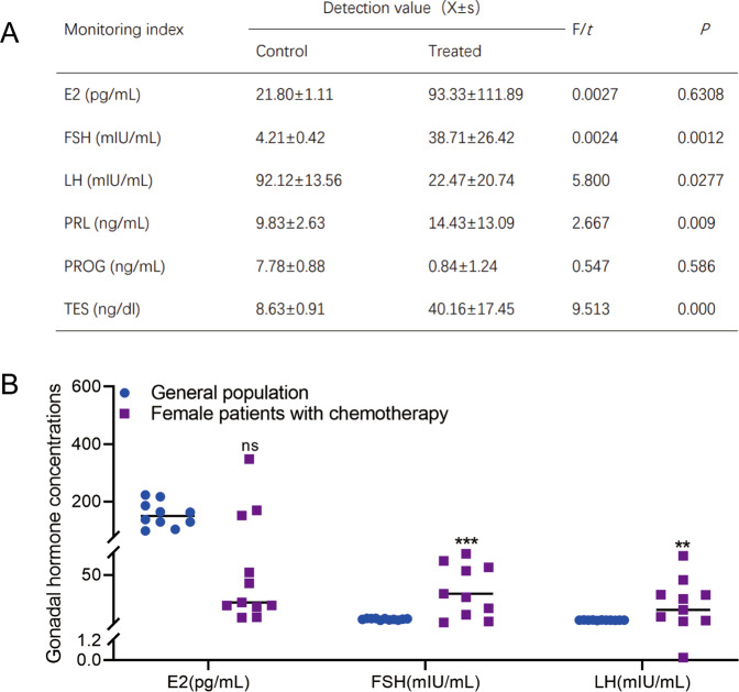
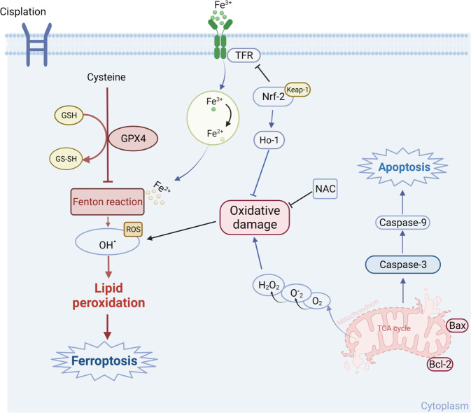
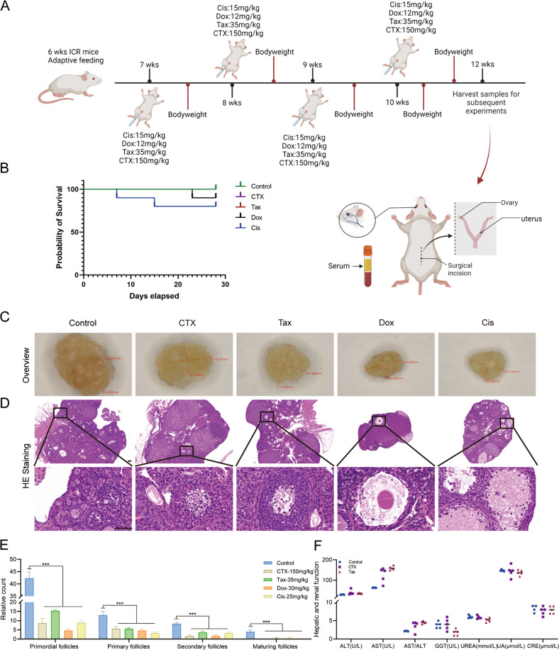
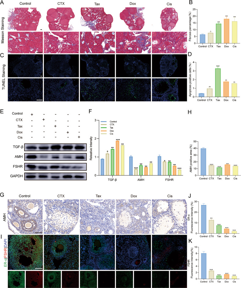
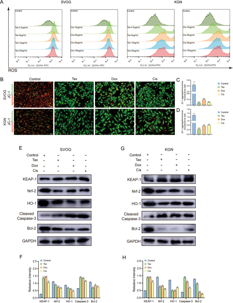
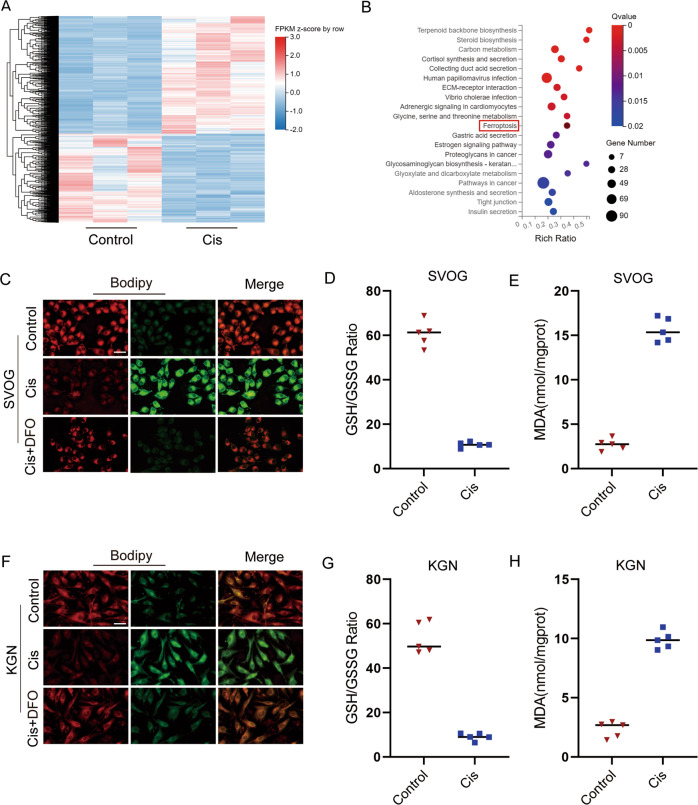
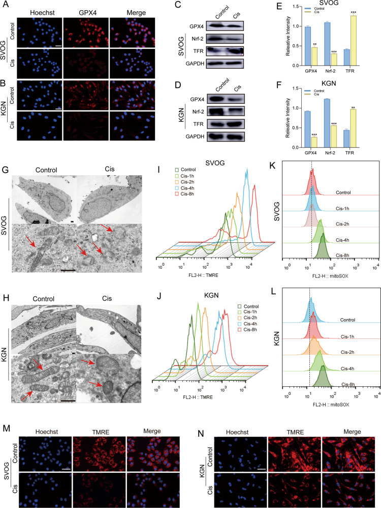
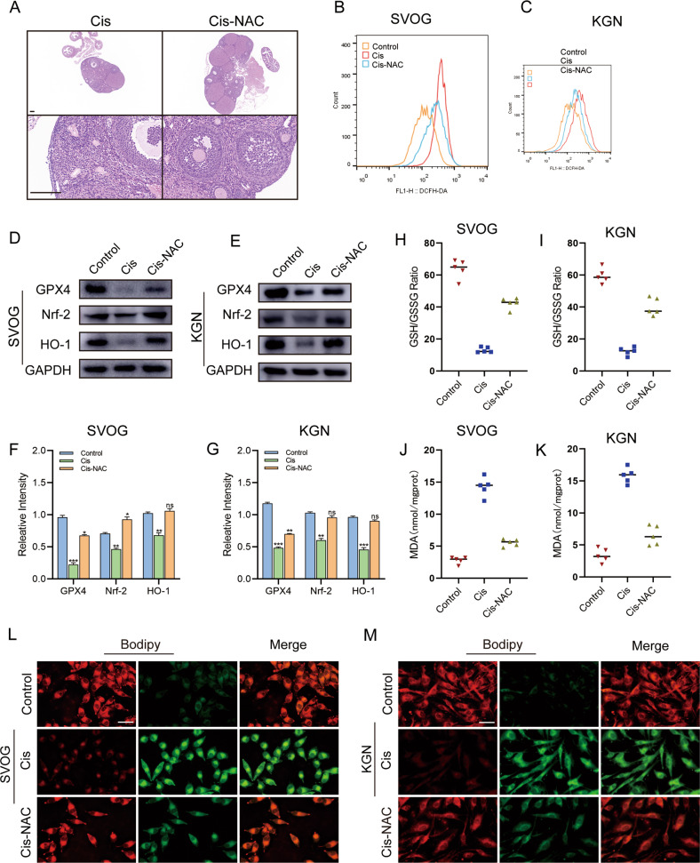
Chemotherapy was conventionally applied to kill cancer cells, but regrettably, they also induce damage to normal cells with high-proliferative capacity resulting in cardiotoxicity, nephrotoxicity, peripheral nerve toxicity, and ovarian toxicity. Of these, chemotherapy-induced ovarian damages mainly include but are not limited to decreased ovarian reserve, infertility, and ovarian atrophy. Therefore, exploring the underlying mechanism of chemotherapeutic drug-induced ovarian damage will pave the way to develop fertility-protective adjuvants for female patients during conventional cancer treatment. Herein, we firstly confirmed the abnormal gonadal hormone levels in patients who received chemotherapy and further found that conventional chemotherapeutic drugs (cyclophosphamide, CTX; paclitaxel, Tax; doxorubicin, Dox and cisplatin, Cis) treatment significantly decreased both the ovarian volume of mice and the number of primordial and antral follicles and accompanied with the ovarian fibrosis and reduced ovarian reserve in animal models. Subsequently, Tax, Dox, and Cis treatment can induce the apoptosis of ovarian granulosa cells (GCs), likely resulting from excessive reactive oxygen species (ROS) production-induced oxidative damage and impaired cellular anti-oxidative capacity. Thirdly, the following experiments demonstrated that Cis treatment could induce mitochondrial dysfunction through overproducing superoxide in GCs and trigger lipid peroxidation leading to ferroptosis, first reported in chemotherapy-induced ovarian damage. In addition, N-acetylcysteine (NAC) treatment could alleviate the Cis-induced toxicity in GCs by downregulating cellular ROS levels and enhancing the anti-oxidative capacity (promoting the expression of glutathione peroxidase, GPX4; nuclear factor erythroid 2-related factor 2, Nrf2 and heme oxygenase-1, HO-1). Our study confirmed the chemotherapy-induced chaotic hormonal state and ovarian damage in preclinical and clinical examination and indicated that chemotherapeutic drugs initiated ferroptosis in ovarian cells through excessive ROS-induced lipid peroxidation and mitochondrial dysfunction, leading to ovarian cell death. Consequently, developing fertility protectants from the chemotherapy-induced oxidative stress and ferroptosis perspective will ameliorate ovarian damage and further improve the life quality of cancer patients.
化疗通常用于杀死癌细胞,但遗憾的是,它们也会导致高增殖能力的正常细胞受损,从而导致心脏毒性、肾毒性、周围神经毒性和卵巢毒性。其中,化疗引起的卵巢损伤主要包括但不限于卵巢储备减少、不孕和卵巢萎缩。因此,探索化疗药物引起卵巢损伤的潜在机制将为女性患者在常规癌症治疗中开发生育保护辅助剂铺平道路。在这里,我们首先证实了接受化疗的患者存在异常的性腺激素水平,进一步发现常规化疗药物(环磷酰胺、CTX;紫杉醇、Tax;多柔比星、Dox 和顺铂、Cis)治疗显著降低了小鼠的卵巢体积和原始卵泡和窦卵泡的数量,并伴有卵巢纤维化和动物模型中卵巢储备减少。随后,Tax、Dox 和 Cis 治疗可诱导卵巢颗粒细胞(GCs)凋亡,可能是由于活性氧(ROS)产生过多引起的氧化损伤和细胞抗氧化能力受损所致。第三,实验表明 Cis 处理可通过在 GCs 中超产超氧化物引起线粒体功能障碍,并引发脂质过氧化导致铁死亡,这是在化疗引起的卵巢损伤中首次报道的。此外,N-乙酰半胱氨酸(NAC)处理可通过下调细胞 ROS 水平和增强抗氧化能力(促进谷胱甘肽过氧化物酶、GPX4;核因子红细胞 2 相关因子 2、Nrf2 和血红素加氧酶-1、HO-1 的表达)来缓解 Cis 对 GCs 的毒性。我们的研究在临床前和临床检查中证实了化疗引起的激素紊乱和卵巢损伤,并表明化疗药物通过过量 ROS 诱导的脂质过氧化和线粒体功能障碍引发卵巢细胞中的铁死亡,导致卵巢细胞死亡。因此,从化疗引起的氧化应激和铁死亡的角度开发生育保护剂将改善卵巢损伤,进一步提高癌症患者的生活质量。