• 文献检索
  • 文档翻译
  • 深度研究
  • 学术资讯
  • Suppr Zotero 插件Zotero 插件
  • 邀请有礼
  • 套餐&价格
  • 历史记录
应用&插件
Suppr Zotero 插件Zotero 插件浏览器插件Mac 客户端Windows 客户端微信小程序
定价
高级版会员购买积分包购买API积分包
服务
文献检索文档翻译深度研究API 文档MCP 服务
关于我们
关于 Suppr公司介绍联系我们用户协议隐私条款
关注我们

Suppr 超能文献

核心技术专利:CN118964589B侵权必究
粤ICP备2023148730 号-1Suppr @ 2026

文献检索

告别复杂PubMed语法,用中文像聊天一样搜索,搜遍4000万医学文献。AI智能推荐,让科研检索更轻松。

立即免费搜索

文件翻译

保留排版,准确专业,支持PDF/Word/PPT等文件格式,支持 12+语言互译。

免费翻译文档

深度研究

AI帮你快速写综述,25分钟生成高质量综述,智能提取关键信息,辅助科研写作。

立即免费体验

高眼压症患者降低眼压后视网膜血管密度的变化

Change of Retinal Vessel Density After Lowering Intraocular Pressure in Ocular Hypertension.

作者信息

Chen Xuhao, Hong Ying, Di Haohao, Wu Qianru, Zhang Di, Zhang Chun

机构信息

Department of Ophthalmology, Peking University Third Hospital, Beijing, China.

Beijing Key Laboratory of Restoration of Damaged Ocular Nerve, Peking University Third Hospital, Beijing, China.

出版信息

Front Med (Lausanne). 2021 Dec 9;8:730327. doi: 10.3389/fmed.2021.730327. eCollection 2021.

DOI:10.3389/fmed.2021.730327
PMID:34957136
原文链接:https://pmc.ncbi.nlm.nih.gov/articles/PMC8695549/
Abstract

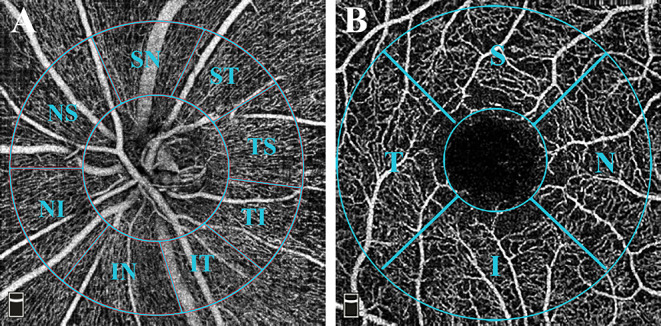
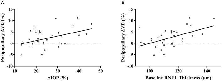
To investigate the relationship between retinal microvasculature changes and intraocular pressure (IOP) for ocular hypertension (OHT) patients and further assess the factors associated with retinal microcirculation changes. This was a single-center prospective study designed for OHT patients, which consisted of two visits. After collecting baseline data of those who met the eligibility criteria, these patients were treated with latanoprost 0.005% ophthalmic solution for 4 weeks. Peripapillary vessel density (VD) of radial peripapillary capillaries (RPC) layer, macular VD in both superficial and deep layers, and foveal avascular zone (FAZ) area were measured by optical coherence tomography angiography (OCTA) before and after the treatment. We compared the changes in IOP and VD among the two visits by paired-sample -test. Bonferroni correction was applied. Factors associated with VD changes were analyzed by linear regression analysis. Thirty-four eyes of thirty-four patients were included. The mean IOP decreased by 6.5 ± 2.2 mmHg ( < 0.001). The peripapillary RPC VD increased significantly from 51.8 ± 2.5 to 53.0 ± 3.1% (Adjusted- = 0.012). We found no significant difference in detailed sectors of the peripapillary region after correction. In the macular area, both the superficial and deep layers in foveal (superficial: 0.2 ± 1.9%, = 0.523; deep: 0.0 ± 2.3%, = 0.969) and parafoveal (superficial: 0.3 ± 3.0%, = 0.565; deep: 0.5 ± 3.1%, = 0.423) VD remained unchanged. The decrease of the mean FAZ area was insignificant ( = 0.295). The percentage of IOP reduction (β = 0.330, = 0.031) and the baseline RNFL thickness (β = 0.450, = 0.004) significantly correlated with the percentage of peripapillary RPC VD improvement in the multivariate linear regression analysis. The peripapillary VD in OHT patients increased after the reduction of IOP. The mild change of IOP did not alter the microcirculation in the macula. In addition, the percentage of IOP change and the baseline RNFL thickness were independent factors for the peripapillary RPC VD improvement.

摘要

为研究高眼压症(OHT)患者视网膜微血管变化与眼压(IOP)之间的关系,并进一步评估与视网膜微循环变化相关的因素。这是一项针对OHT患者的单中心前瞻性研究,包括两次就诊。在收集符合入选标准患者的基线数据后,这些患者使用0.005%拉坦前列素滴眼液治疗4周。通过光学相干断层扫描血管造影(OCTA)测量治疗前后视乳头周围放射状毛细血管(RPC)层的视乳头周围血管密度(VD)、黄斑浅层和深层的黄斑VD以及中心凹无血管区(FAZ)面积。我们通过配对样本t检验比较两次就诊时IOP和VD的变化。应用Bonferroni校正。通过线性回归分析分析与VD变化相关的因素。纳入了34例患者的34只眼。平均IOP降低了6.5±2.2 mmHg(P<0.001)。视乳头周围RPC VD从51.8±2.5%显著增加至53.0±3.1%(校正后P = 0.012)。校正后,我们发现视乳头周围区域的详细扇区无显著差异。在黄斑区,中心凹(浅层:0.2±1.9%,P = 0.523;深层:0.0±2.3%,P = 0.969)和旁中心凹(浅层:0.3±3.0%,P = 0.565;深层:0.5±3.1%,P = 0.423)浅层和深层的VD均无变化。平均FAZ面积的减小不显著(P = 0.295)。在多变量线性回归分析中,IOP降低百分比(β = 0.330,P = 0.031)和基线视网膜神经纤维层(RNFL)厚度(β = 0.450,P = 0.004)与视乳头周围RPC VD改善百分比显著相关。OHT患者视乳头周围VD在IOP降低后增加。IOP的轻微变化未改变黄斑区的微循环。此外,IOP变化百分比和基线RNFL厚度是视乳头周围RPC VD改善的独立因素。

https://cdn.ncbi.nlm.nih.gov/pmc/blobs/9815/8695549/b2260b6f9354/fmed-08-730327-g0002.jpg
https://cdn.ncbi.nlm.nih.gov/pmc/blobs/9815/8695549/3a0b11a9d490/fmed-08-730327-g0001.jpg
https://cdn.ncbi.nlm.nih.gov/pmc/blobs/9815/8695549/b2260b6f9354/fmed-08-730327-g0002.jpg
https://cdn.ncbi.nlm.nih.gov/pmc/blobs/9815/8695549/3a0b11a9d490/fmed-08-730327-g0001.jpg
https://cdn.ncbi.nlm.nih.gov/pmc/blobs/9815/8695549/b2260b6f9354/fmed-08-730327-g0002.jpg

相似文献

1
Change of Retinal Vessel Density After Lowering Intraocular Pressure in Ocular Hypertension.高眼压症患者降低眼压后视网膜血管密度的变化
Front Med (Lausanne). 2021 Dec 9;8:730327. doi: 10.3389/fmed.2021.730327. eCollection 2021.
2
Quantitative Analysis of Microvasculature in Macular and Peripapillary Regions in Early Primary Open-Angle Glaucoma.早期原发性开角型青光眼黄斑及视盘周围区域微血管定量分析。
Curr Eye Res. 2020 May;45(5):629-635. doi: 10.1080/02713683.2019.1676912. Epub 2019 Oct 14.
3
The evaluation of juvenile ocular hypertension by optical coherence tomography angiography.光学相干断层扫描血管造影术对青少年高眼压症的评估
BMC Ophthalmol. 2020 Oct 21;20(1):423. doi: 10.1186/s12886-020-01641-4.
4
Effect of surgical intraocular pressure lowering on retinal structures - nerve fibre layer, foveal avascular zone, peripapillary and macular vessel density: 1 year results.手术降眼压对视网膜结构的影响——神经纤维层、黄斑中心无血管区、视盘周围和黄斑血管密度:1 年结果。
Eye (Lond). 2020 Mar;34(3):562-571. doi: 10.1038/s41433-019-0560-6. Epub 2019 Aug 13.
5
Changes in peripapillary and macular vascular density after laser selective trabeculoplasty: an optical coherence tomography angiography study.激光选择性小梁成形术后视乳头周围和黄斑区血管密度的变化:一项光学相干断层扫描血管造影研究
Acta Ophthalmol. 2022 Mar;100(2):203-211. doi: 10.1111/aos.14805. Epub 2021 Feb 24.
6
The Effect of Medical Lowering of Intraocular Pressure on Peripapillary and Macular Blood Flow as Measured by Optical Coherence Tomography Angiography in Treatment-naive Eyes.未经治疗的眼内压低对光学相干断层扫描血管造影测量的视盘和黄斑血流的影响。
J Glaucoma. 2021 Jun 1;30(6):465-472. doi: 10.1097/IJG.0000000000001828.
7
Evaluation of macular and peripapillary vascular parameter change in healthy subjects after caffeine intake using optical coherence tomography angiography.评估健康受试者摄入咖啡因后利用光相干断层扫描血管造影术的黄斑和视盘周围血管参数的变化。
Indian J Ophthalmol. 2022 Mar;70(3):879-889. doi: 10.4103/ijo.IJO_1989_21.
8
Immediate Changes in Peripapillary Retinal Vasculature after Intraocular Pressure Elevation -an Optical Coherence Tomography Angiography Study.眼压升高后视乳头周围视网膜血管的即时变化——一项光相干断层扫描血管造影研究。
Curr Eye Res. 2020 Jun;45(6):749-756. doi: 10.1080/02713683.2019.1695843. Epub 2019 Nov 27.
9
Optical coherence tomography angiography of the peripapillary region and macula in normal, primary open angle glaucoma, pseudoexfoliation glaucoma and ocular hypertension eyes.正常眼、原发性开角型青光眼、剥脱性青光眼和高眼压症患者眼的视乳头周围区域和黄斑的光学相干断层扫描血管造影
Int J Ophthalmol. 2020 May 18;13(5):744-754. doi: 10.18240/ijo.2020.05.08. eCollection 2020.
10
Comparison of vessel density in macular and peripapillary regions between primary open-angle glaucoma and pseudoexfoliation glaucoma using OCTA.应用 OCTA 比较原发性开角型青光眼和假性剥脱综合征患者黄斑区和视盘周围血管密度。
Int Ophthalmol. 2021 Jan;41(1):173-184. doi: 10.1007/s10792-020-01564-5. Epub 2020 Aug 26.

引用本文的文献

1
Vascular Density and Retinal Structure Relationships Across Sectors and Regions of the Optic Nerve in Patients With Primary Open Angle Glaucoma.原发性开角型青光眼患者视神经各扇区和区域的血管密度与视网膜结构关系
J Glaucoma. 2025 Apr 1;34(4):267-274. doi: 10.1097/IJG.0000000000002536. Epub 2025 Jan 16.
2
The effects of topical latanoprost 0.005% treatment on microvascular changes in the optic nerve head and macula.局部应用拉坦前列素 0.005%对视盘和黄斑的微血管变化的影响。
Indian J Ophthalmol. 2024 Nov 1;72(Suppl 5):S907-S912. doi: 10.4103/IJO.IJO_648_24. Epub 2024 Oct 25.
3
The impact of intraocular pressure on optical coherence tomography angiography: A review of current evidence.

本文引用的文献

1
Juvenile-onset open-angle glaucoma - A clinical and genetic update.青少年型开角型青光眼——临床与遗传学更新。
Surv Ophthalmol. 2022 Jul-Aug;67(4):1099-1117. doi: 10.1016/j.survophthal.2021.09.001. Epub 2021 Sep 16.
2
Age and signal strength-related changes in vessel density in the choroid and the retina: an OCT angiography study of the macula and optic disc.年龄与脉络膜和视网膜血管密度的信号强度相关性变化:黄斑和视盘的 OCT 血管造影研究。
Acta Ophthalmol. 2022 Aug;100(5):e1095-e1102. doi: 10.1111/aos.15028. Epub 2021 Sep 16.
3
Longer Axial Length Potentiates Relationship of Intraocular Pressure and Peripapillary Vessel Density in Glaucoma Patients.
眼压对光学相干断层扫描血管造影的影响:当前证据综述
Saudi J Ophthalmol. 2024 Jan 3;38(2):144-151. doi: 10.4103/sjopt.sjopt_112_23. eCollection 2024 Apr-Jun.
4
Functional and perfusion changes associated with silicone oil tamponade after macula-off rhegmatogenous retinal detachment surgery: an optical coherence tomography angiography/microperimetry study.硅油填充术后黄斑裂孔性视网膜脱离复位术后与功能和灌注变化相关:光学相干断层扫描血管造影/微视野研究。
Int Ophthalmol. 2024 Feb 22;44(1):107. doi: 10.1007/s10792-024-03037-5.
5
Peripapillary Vascular Density in Compressive Optic Neuropathy and Normal-Tension Glaucoma: A Severity-Controlled Comparison.压迫性视神经病变和正常眼压性青光眼的视盘旁血管密度:严重程度对照比较。
Invest Ophthalmol Vis Sci. 2023 Sep 1;64(12):10. doi: 10.1167/iovs.64.12.10.
6
A comparison of peripapillary vessel density between subjects with normal-tension glaucoma and primary open-angle glaucoma with similar extents of glaucomatous damage.正常眼压性青光眼和原发性开角型青光眼患者的视盘周围血管密度比较,这些患者的青光眼损害程度相似。
Sci Rep. 2023 Jun 7;13(1):9258. doi: 10.1038/s41598-023-36369-w.
7
Axons of retinal ganglion cells on the optic nerve disc following vessel density correction at different IOP values.在不同眼压值下进行血管密度校正后,视神经盘上视网膜神经节细胞的轴突。
Exp Ther Med. 2023 Apr 19;25(6):261. doi: 10.3892/etm.2023.11960. eCollection 2023 Jun.
眼轴长度与青光眼患者眼压和视盘周围血管密度的关系。
Invest Ophthalmol Vis Sci. 2021 Jul 1;62(9):37. doi: 10.1167/iovs.62.9.37.
4
The Effect of Medical Lowering of Intraocular Pressure on Peripapillary and Macular Blood Flow as Measured by Optical Coherence Tomography Angiography in Treatment-naive Eyes.未经治疗的眼内压低对光学相干断层扫描血管造影测量的视盘和黄斑血流的影响。
J Glaucoma. 2021 Jun 1;30(6):465-472. doi: 10.1097/IJG.0000000000001828.
5
OCT-angiography detects longitudinal microvascular changes in glaucoma: a systematic review.光学相干断层扫描血管造影术检测青光眼纵向微血管变化:一项系统评价
Br J Ophthalmol. 2022 May;106(5):667-675. doi: 10.1136/bjophthalmol-2020-318166. Epub 2021 Jan 15.
6
Recent developments in the use of optical coherence tomography for glaucoma.光学相干断层扫描在青光眼应用中的新进展。
Curr Opin Ophthalmol. 2021 Mar 1;32(2):98-104. doi: 10.1097/ICU.0000000000000733.
7
OCT angiography analysis of retinal vessel density in primary open-angle glaucoma with and without Tafluprost therapy.原发性开角型青光眼患者使用他氟前列素治疗与未使用治疗时视网膜血管密度的光学相干断层扫描血管造影分析
BMC Ophthalmol. 2020 Nov 12;20(1):444. doi: 10.1186/s12886-020-01707-3.
8
OCT angiography measured changes in the foveal avascular zone area after glaucoma surgery.OCT 血管造影测量青光眼手术后中心凹无血管区面积的变化。
Br J Ophthalmol. 2022 Jan;106(1):80-86. doi: 10.1136/bjophthalmol-2020-317038. Epub 2020 Nov 5.
9
Myopia-related stepwise and quadrant retinal microvascular alteration and its correlation with axial length.近视相关的阶梯状和象限性视网膜微血管改变及其与眼轴长度的相关性。
Eye (Lond). 2021 Aug;35(8):2196-2205. doi: 10.1038/s41433-020-01225-y. Epub 2020 Oct 21.
10
The evaluation of juvenile ocular hypertension by optical coherence tomography angiography.光学相干断层扫描血管造影术对青少年高眼压症的评估
BMC Ophthalmol. 2020 Oct 21;20(1):423. doi: 10.1186/s12886-020-01641-4.