Department of Prosthodontics, Affiliated Stomatology Hospital of Guangzhou Medical University, Guangzhou, People's Republic of China.
Guangdong Engineering Research Center of Oral Restoration and Reconstruction, Guangzhou Key Laboratory of Basic and Applied Research of Oral Regenerative Medicine, Guangzhou, People's Republic of China.
Int J Nanomedicine. 2023 Jun 13;18:3141-3155. doi: 10.2147/IJN.S404047. eCollection 2023.
Improving the biological sealing around dental abutments could promote the long-term success of implants. Although titanium abutments have a wide range of clinical applications, they incur esthetic risks due to their color, especially in the esthetic zone. Currently, zirconia has been applied as an esthetic alternative material for implant abutments; however, zirconia is purported to be an inert biomaterial. How to improve the biological activities of zirconia has thus become a popular research topic. In this study, we presented a novel self-glazed zirconia (SZ) surface with nanotopography fabricated by additive 3D gel deposition and investigated its soft tissue integration capability compared to that of clinically used titanium and polished conventional zirconia surfaces.
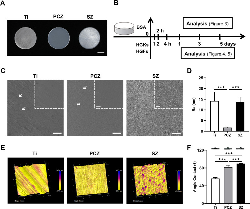
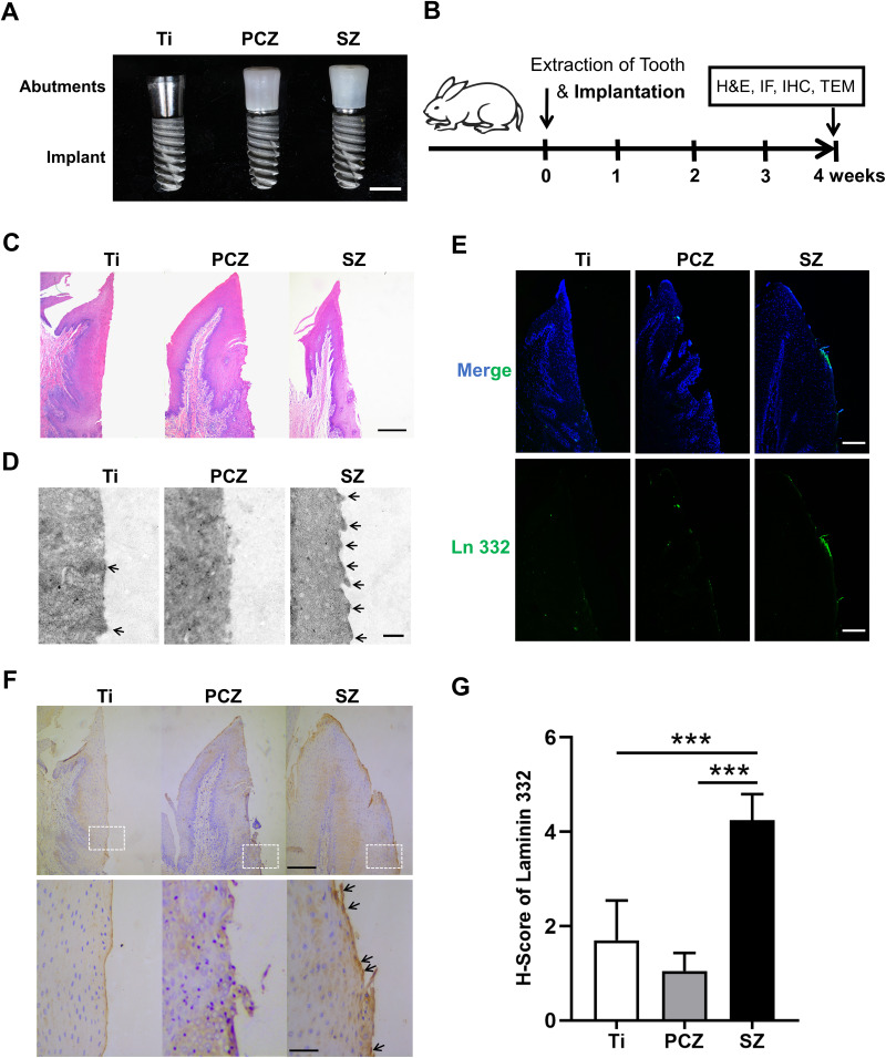
Three groups of disc samples were prepared for in vitro study and the three groups of abutment samples were prepared for in vivo study. The surface topography, roughness, wettability and chemical composition of the samples were examined. Moreover, we analyzed the effect of the three groups of samples on protein adsorption and on the biological behavior of human gingival keratinocytes (HGKs) and human gingival fibroblasts (HGFs). Furthermore, we conducted an in vivo study in which the bilateral mandibular anterior teeth of rabbits were extracted and replaced with implants and corresponding abutments.
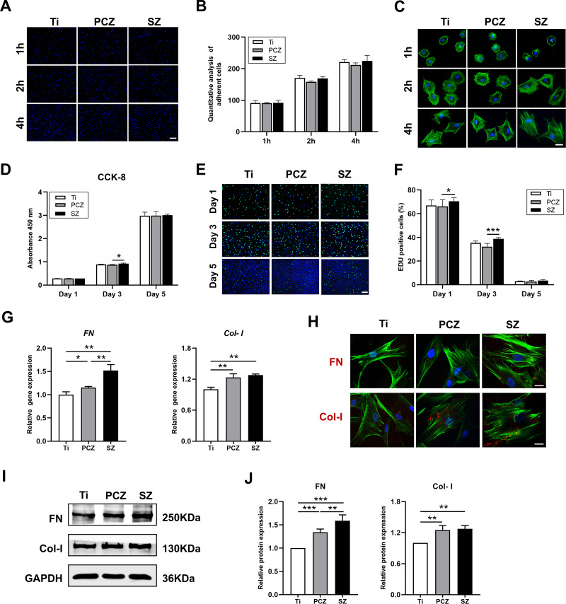
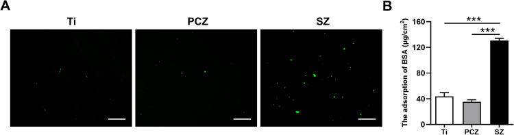
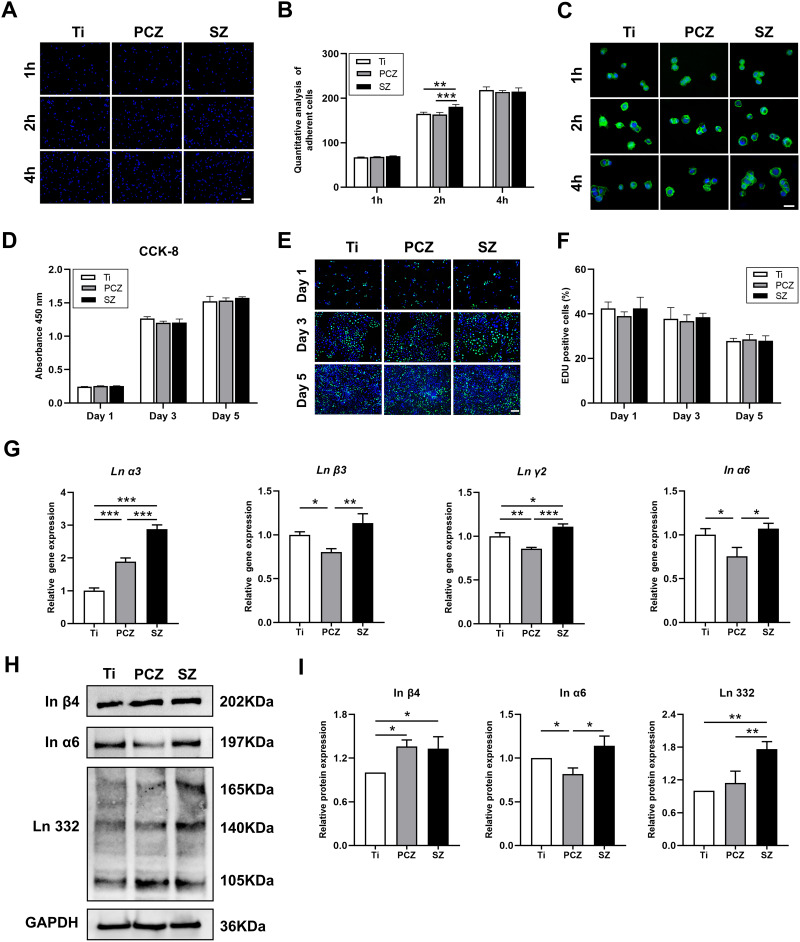
The surface of SZ showed a unique nanotopography with nm range roughness and a greater ability to absorb protein. The promoted expression of adhesion molecules in both HGKs and HGFs was observed on the SZ surface compared to the surfaces of Ti and PCZ, while the cell viability and proliferation of HGKs and the number of HGFs adhesion were not significant among all groups. In vivo results showed that the SZ abutment formed strong biological sealing at the abutment-soft tissue interface and exhibited markedly more hemidesmosomes when observed with a transmission electron microscope.
These results demonstrated that the novel SZ surface with nanotopography promoted soft tissue integration, suggesting its promising application as a zirconia surface for the dental abutment.
改善牙基台周围的生物密封性可以提高种植体的长期成功率。尽管钛基台具有广泛的临床应用,但由于其颜色,尤其是在美学区域,存在美学风险。目前,氧化锆已被用作种植体基台的美学替代材料;然而,氧化锆被认为是一种惰性生物材料。因此,如何提高氧化锆的生物活性已成为一个热门研究课题。本研究提出了一种具有纳米形貌的新型自上釉氧化锆(SZ)表面,该表面采用添加剂 3D 凝胶沉积技术制备,并对其与临床应用的钛和抛光常规氧化锆表面的软组织整合能力进行了研究。
本研究进行了体外和体内实验。体外实验制备了三组圆盘样品,体内实验制备了三组基台样品。对样品的表面形貌、粗糙度、润湿性和化学成分进行了检测。此外,还分析了三组样品对蛋白质吸附和人牙龈角质细胞(HGKs)和人牙龈成纤维细胞(HGFs)生物学行为的影响。此外,还进行了一项体内研究,在该研究中,兔子的双侧下颌前牙被拔出并用种植体和相应的基台替代。
SZ 表面呈现独特的纳米形貌,具有纳米级粗糙度,且具有更强的蛋白质吸附能力。与 Ti 和 PCZ 表面相比,SZ 表面促进了 HGKs 和 HGFs 中黏附分子的表达,而 HGKs 的细胞活力和增殖以及 HGFs 的黏附数量在各组之间没有显著差异。体内结果表明,SZ 基台在基台-软组织界面形成了强有力的生物密封,用透射电子显微镜观察时,明显有更多的半桥粒。
这些结果表明,具有纳米形貌的新型 SZ 表面促进了软组织整合,表明其有望作为牙基台用氧化锆表面的应用。