Department of Electronics and Communication EngineeringNational Institute of Technology at Calicut Kozhikode 673601 India.
Department of Computer ScienceNorwegian University of Science and Technology 7034 Trondheim Norway.
IEEE J Transl Eng Health Med. 2023 Jun 6;11:360-374. doi: 10.1109/JTEHM.2023.3283444. eCollection 2023.
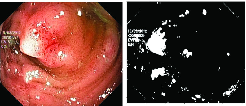
Endoscopy is a medical diagnostic procedure used to see inside the human body with the help of a camera-attached system called the endoscope. Endoscopic images and videos suffer from specular reflections (or highlight) and can have an adverse impact on the diagnostic quality of images. These scattered white regions severely affect the visual appearance of images for both endoscopists and the computer-aided diagnosis of diseases. Methods & Results: We introduce a new parameter-free matrix decomposition technique to remove the specular reflections. The proposed method decomposes the original image into a highlight-free pseudo-low-rank component and a highlight component. Along with the highlight removal, the approach also removes the boundary artifacts present around the highlight regions, unlike the previous works based on family of Robust Principal Component Analysis (RPCA). The approach is evaluated on three publicly available endoscopy datasets: Kvasir Polyp, Kvasir Normal-Pylorus and Kvasir Capsule datasets. Our evaluation is benchmarked against 4 different state-of-the-art approaches using three different well-used metrics such as Structural Similarity Index Measure (SSIM), Percentage of highlights remaining and Coefficient of Variation (CoV). Conclusions: The results show significant improvements over the compared methods on all three metrics. The approach is further validated for statistical significance where it emerges better than other state-of-the-art approaches.The mathematical concepts of low rank and rank decomposition in matrix algebra are translated to remove specularities in the endoscopic images The result shows the impact of the proposed method in removing specular reflections from endoscopic images indicating improved diagnosis efficiency for both endoscopists and computer-aided diagnosis systems.
内窥镜检查是一种医学诊断程序,借助称为内窥镜的相机系统来观察人体内部。内窥镜图像和视频会受到镜面反射(或高光)的影响,这会对图像的诊断质量产生不利影响。这些散射的白色区域严重影响了内窥镜医生和疾病计算机辅助诊断对图像的视觉外观。
我们引入了一种新的无参数矩阵分解技术来去除镜面反射。该方法将原始图像分解为无高光的伪低秩分量和高光分量。与基于鲁棒主成分分析(RPCA)族的先前方法不同,该方法不仅可以去除高光区域周围的边界伪影,还可以去除高光。该方法在三个公开可用的内窥镜数据集上进行了评估:Kvasir Polyp、Kvasir Normal-Pylorus 和 Kvasir Capsule 数据集。我们使用三种不同的常用指标,如结构相似性指数度量(SSIM)、高光残留百分比和变化系数(CoV),对 4 种不同的最先进方法进行了评估。
与比较方法相比,该方法在所有三个指标上都有显著的改进。该方法还针对统计学意义进行了验证,结果表明其优于其他最先进的方法。矩阵代数中的低秩和秩分解的数学概念被转化为去除内窥镜图像中的镜面反射。结果表明,该方法在去除内窥镜图像中的镜面反射方面具有显著效果,这将提高内窥镜医生和计算机辅助诊断系统的诊断效率。