Centre for the Developing Brain, School of Biomedical Engineering & Imaging Sciences, King's College London, London, UK.
Biomedical Engineering Department, School of Biomedical Engineering & Imaging Sciences, King's College London, London, UK.
Magn Reson Med. 2023 Dec;90(6):2306-2320. doi: 10.1002/mrm.29803. Epub 2023 Jul 19.
To improve motion robustness of functional fetal MRI scans by developing an intrinsic real-time motion correction method. MRI provides an ideal tool to characterize fetal brain development and growth. It is, however, a relatively slow imaging technique and therefore extremely susceptible to subject motion, particularly in functional MRI experiments acquiring multiple Echo-Planar-Imaging-based repetitions, for example, diffusion MRI or blood-oxygen-level-dependency MRI.
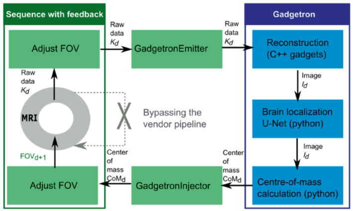
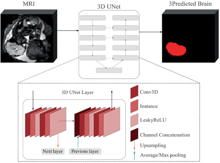
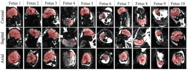
A 3D UNet was trained on 125 fetal datasets to track the fetal brain position in each repetition of the scan in real time. This tracking, inserted into a Gadgetron pipeline on a clinical scanner, allows updating the position of the field of view in a modified echo-planar imaging sequence. The method was evaluated in real-time in controlled-motion phantom experiments and ten fetal MR studies (17 + 4-34 + 3 gestational weeks) at 3T. The localization network was additionally tested retrospectively on 29 low-field (0.55T) datasets.
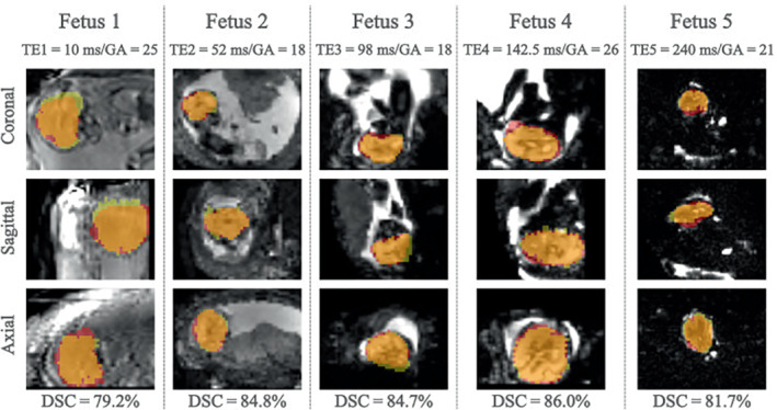
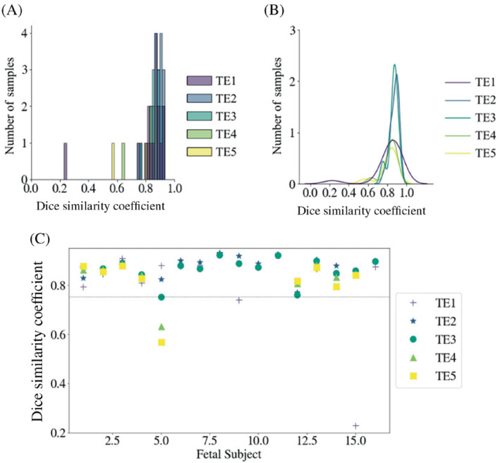
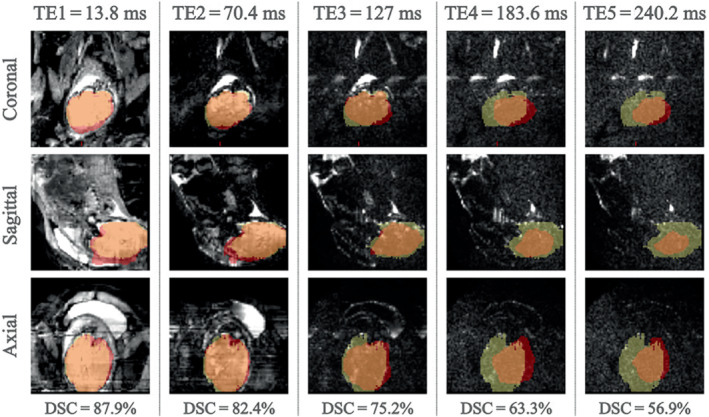
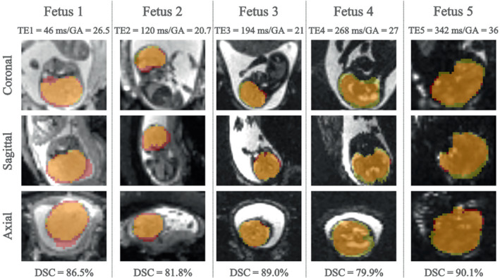
Our method achieved real-time fetal head tracking and prospective correction of the acquisition geometry. Localization performance achieved Dice scores of 84.4% and 82.3%, respectively for both the unseen 1.5T/3T and 0.55T fetal data, with values higher for cephalic fetuses and increasing with gestational age.
Our technique was able to follow the fetal brain even for fetuses under 18 weeks GA in real-time at 3T and was successfully applied "offline" to new cohorts on 0.55T. Next, it will be deployed to other modalities such as fetal diffusion MRI and to cohorts of pregnant participants diagnosed with pregnancy complications, for example, pre-eclampsia and congenital heart disease.
通过开发一种内在的实时运动校正方法,提高功能胎儿 MRI 扫描的运动鲁棒性。MRI 提供了一种理想的工具来描述胎儿大脑的发育和生长。然而,它是一种相对较慢的成像技术,因此非常容易受到受试者运动的影响,特别是在功能 MRI 实验中,需要获取多个基于回波平面成像的重复,例如扩散 MRI 或血氧水平依赖性 MRI。
在 125 个胎儿数据集上训练一个 3D UNet,以实时跟踪扫描中每个重复的胎儿大脑位置。这种跟踪被插入到临床扫描仪上的 Gadgetron 管道中,可以在修改后的回波平面成像序列中更新视场的位置。该方法在受控运动体模实验和 10 项胎儿磁共振研究(17+4-34+3 孕周)中进行了实时评估。该定位网络还在 29 项低场(0.55T)数据集上进行了回顾性测试。
我们的方法实现了实时胎儿头部跟踪和前瞻性采集几何校正。定位性能在未见的 1.5T/3T 和 0.55T 胎儿数据中分别达到了 84.4%和 82.3%的 Dice 评分,对于头部胎儿更高,并且随着胎龄的增加而增加。
我们的技术即使在 3T 下,也能够实时跟踪胎儿大脑,并且在 0.55T 上成功应用于新的队列。下一步,它将被部署到其他模态,如胎儿扩散 MRI,并应用于患有妊娠并发症(如子痫前期和先天性心脏病)的孕妇队列。