• 文献检索
  • 文档翻译
  • 深度研究
  • 学术资讯
  • Suppr Zotero 插件Zotero 插件
  • 邀请有礼
  • 套餐&价格
  • 历史记录
应用&插件
Suppr Zotero 插件Zotero 插件浏览器插件Mac 客户端Windows 客户端微信小程序
定价
高级版会员购买积分包购买API积分包
服务
文献检索文档翻译深度研究API 文档MCP 服务
关于我们
关于 Suppr公司介绍联系我们用户协议隐私条款
关注我们

Suppr 超能文献

核心技术专利:CN118964589B侵权必究
粤ICP备2023148730 号-1Suppr @ 2026

文献检索

告别复杂PubMed语法,用中文像聊天一样搜索,搜遍4000万医学文献。AI智能推荐,让科研检索更轻松。

立即免费搜索

文件翻译

保留排版,准确专业,支持PDF/Word/PPT等文件格式,支持 12+语言互译。

免费翻译文档

深度研究

AI帮你快速写综述,25分钟生成高质量综述,智能提取关键信息,辅助科研写作。

立即免费体验

利用深度学习技术自动检测和分割 CT 肺动脉造影(CTPA)中的肺栓塞,无需手动勾画。

Automated detection and segmentation of pulmonary embolisms on computed tomography pulmonary angiography (CTPA) using deep learning but without manual outlining.

机构信息

Department of Radiology, University of Pittsburgh School of Medicine, Pittsburgh, PA 15213, USA; Department of Bioengineering, University of Pittsburgh, Pittsburgh, PA 15213, USA.

Department of Radiology, Dokuz Eylul University, Izmir, Turkey.

出版信息

Med Image Anal. 2023 Oct;89:102882. doi: 10.1016/j.media.2023.102882. Epub 2023 Jul 14.

DOI:10.1016/j.media.2023.102882
PMID:37482032
原文链接:https://pmc.ncbi.nlm.nih.gov/articles/PMC10528048/
Abstract

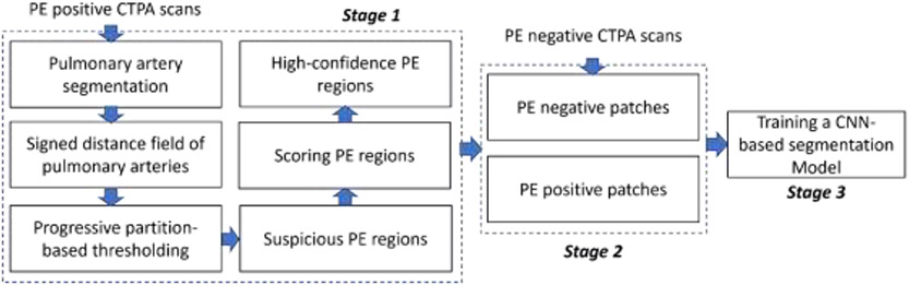
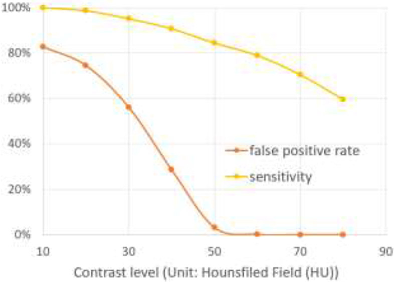
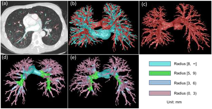
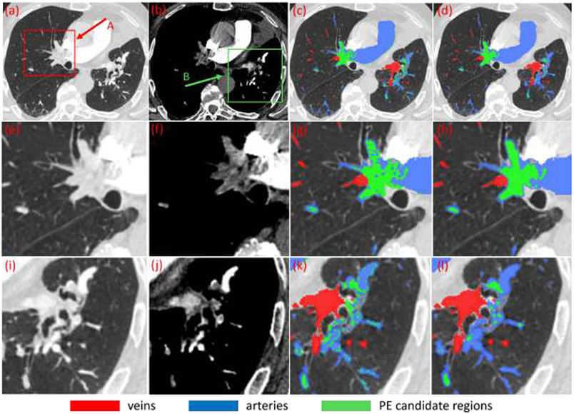
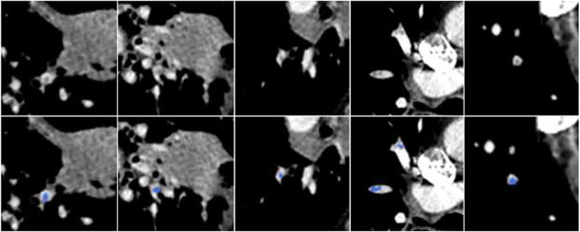
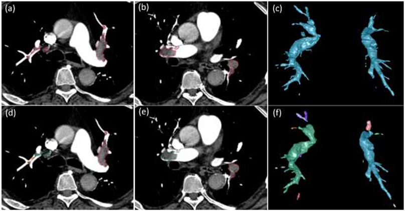
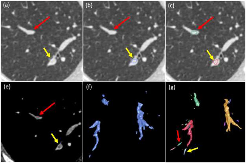
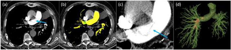
We present a novel computer algorithm to automatically detect and segment pulmonary embolisms (PEs) on computed tomography pulmonary angiography (CTPA). This algorithm is based on deep learning but does not require manual outlines of the PE regions. Given a CTPA scan, both intra- and extra-pulmonary arteries were firstly segmented. The arteries were then partitioned into several parts based on size (radius). Adaptive thresholding and constrained morphological operations were used to identify suspicious PE regions within each part. The confidence of a suspicious region to be PE was scored based on its contrast in the arteries. This approach was applied to the publicly available RSNA Pulmonary Embolism CT Dataset (RSNA-PE) to identify three-dimensional (3-D) PE negative and positive image patches, which were used to train a 3-D Recurrent Residual U-Net (R2-Unet) to automatically segment PE. The feasibility of this computer algorithm was validated on an independent test set consisting of 91 CTPA scans acquired from a different medical institute, where the PE regions were manually located and outlined by a thoracic radiologist (>18 years' experience). An R2-Unet model was also trained and validated on the manual outlines using a 5-fold cross-validation method. The CNN model trained on the high-confident PE regions showed a Dice coefficient of 0.676±0.168 and a false positive rate of 1.86 per CT scan, while the CNN model trained on the manual outlines demonstrated a Dice coefficient of 0.647±0.192 and a false positive rate of 4.20 per CT scan. The former model performed significantly better than the latter model (p<0.01). The promising performance of the developed PE detection and segmentation algorithm suggests the feasibility of training a deep learning network without dedicating significant efforts to manual annotations of the PE regions on CTPA scans.

摘要

我们提出了一种新的计算机算法,用于在计算机断层肺动脉造影(CTPA)上自动检测和分割肺栓塞(PE)。该算法基于深度学习,但不需要手动勾勒 PE 区域。给定 CTPA 扫描,首先对肺内和肺外动脉进行分割。然后,根据大小(半径)将动脉分成几部分。自适应阈值和约束形态学操作用于在每个部分内识别可疑的 PE 区域。根据动脉中的对比度对可疑区域是否为 PE 进行评分。该方法应用于公开的 RSNA 肺栓塞 CT 数据集(RSNA-PE),以识别三维(3-D)PE 阴性和阳性图像补丁,这些补丁用于训练 3-D 递归残差 U-Net(R2-Unet)以自动分割 PE。该计算机算法的可行性在一个由另一家医疗机构获得的 91 例 CTPA 扫描组成的独立测试集中得到验证,其中由一位胸部放射科医生(>18 年经验)手动定位和勾勒出 PE 区域。还使用 5 折交叉验证方法在手动轮廓上训练和验证了 R2-Unet 模型。在高置信度 PE 区域上训练的 CNN 模型的 Dice 系数为 0.676±0.168,假阳性率为每 CT 扫描 1.86,而在手动轮廓上训练的 CNN 模型的 Dice 系数为 0.647±0.192,假阳性率为每 CT 扫描 4.20。前者模型的性能明显优于后者模型(p<0.01)。开发的 PE 检测和分割算法的良好性能表明,在 CTPA 扫描上不需要大量人工标注 PE 区域的情况下,训练深度学习网络是可行的。

https://cdn.ncbi.nlm.nih.gov/pmc/blobs/b128/10528048/e21b5a864ffe/nihms-1919460-f0009.jpg
https://cdn.ncbi.nlm.nih.gov/pmc/blobs/b128/10528048/bfde2f625d60/nihms-1919460-f0002.jpg
https://cdn.ncbi.nlm.nih.gov/pmc/blobs/b128/10528048/5eac3a23f168/nihms-1919460-f0003.jpg
https://cdn.ncbi.nlm.nih.gov/pmc/blobs/b128/10528048/574da0d300e6/nihms-1919460-f0004.jpg
https://cdn.ncbi.nlm.nih.gov/pmc/blobs/b128/10528048/0cd95f0c8b99/nihms-1919460-f0005.jpg
https://cdn.ncbi.nlm.nih.gov/pmc/blobs/b128/10528048/ce9a48bc596a/nihms-1919460-f0006.jpg
https://cdn.ncbi.nlm.nih.gov/pmc/blobs/b128/10528048/817c26f62ec1/nihms-1919460-f0007.jpg
https://cdn.ncbi.nlm.nih.gov/pmc/blobs/b128/10528048/95fa98b23e46/nihms-1919460-f0008.jpg
https://cdn.ncbi.nlm.nih.gov/pmc/blobs/b128/10528048/e21b5a864ffe/nihms-1919460-f0009.jpg

相似文献

1
Automated detection and segmentation of pulmonary embolisms on computed tomography pulmonary angiography (CTPA) using deep learning but without manual outlining.利用深度学习技术自动检测和分割 CT 肺动脉造影(CTPA)中的肺栓塞,无需手动勾画。
Med Image Anal. 2023 Oct;89:102882. doi: 10.1016/j.media.2023.102882. Epub 2023 Jul 14.
2
Computer-aided detection of pulmonary embolism in computed tomographic pulmonary angiography (CTPA): performance evaluation with independent data sets.计算机断层扫描肺动脉造影(CTPA)中肺栓塞的计算机辅助检测:使用独立数据集进行性能评估
Med Phys. 2009 Aug;36(8):3385-96. doi: 10.1118/1.3157102.
3
Automatic segmentation of arterial tree from 3D computed tomographic pulmonary angiography (CTPA) scans.从三维计算机断层肺动脉造影 (CTPA) 扫描中自动分割动脉树。
Comput Assist Surg (Abingdon). 2019 Oct;24(sup2):79-86. doi: 10.1080/24699322.2019.1649077. Epub 2019 Aug 10.
4
A deep learning approach for automated diagnosis of pulmonary embolism on computed tomographic pulmonary angiography.深度学习在计算机断层肺动脉造影诊断肺栓塞中的应用。
BMC Med Imaging. 2022 Nov 11;22(1):195. doi: 10.1186/s12880-022-00916-0.
5
Automatic multiscale enhancement and segmentation of pulmonary vessels in CT pulmonary angiography images for CAD applications.用于CAD应用的CT肺血管造影图像中肺血管的自动多尺度增强与分割
Med Phys. 2007 Dec;34(12):4567-77. doi: 10.1118/1.2804558.
6
Preliminary investigation of computer-aided detection of pulmonary embolism in three-dimensional computed tomography pulmonary angiography images.三维计算机断层扫描肺动脉造影图像中肺栓塞计算机辅助检测的初步研究。
Acad Radiol. 2005 Jun;12(6):782-92. doi: 10.1016/j.acra.2005.01.014.
7
Detection and severity quantification of pulmonary embolism with 3D CT data using an automated deep learning-based artificial solution.使用基于深度学习的自动化人工解决方案对3D CT数据进行肺栓塞的检测和严重程度量化。
Diagn Interv Imaging. 2024 Mar;105(3):97-103. doi: 10.1016/j.diii.2023.09.006. Epub 2023 Oct 11.
8
Seeking an optimal approach for Computer-aided Diagnosis of Pulmonary Embolism.寻求肺栓塞计算机辅助诊断的最佳方法。
Med Image Anal. 2024 Jan;91:102988. doi: 10.1016/j.media.2023.102988. Epub 2023 Oct 13.
9
[Segmentation of acute pulmonary embolism in computed tomography pulmonary angiography using the deep learning method].[基于深度学习方法的计算机断层扫描肺动脉造影中急性肺栓塞的分割]
Tuberk Toraks. 2023 Jun;71(2):131-137. doi: 10.5578/tt.20239916.
10
Anatomically aware dual-hop learning for pulmonary embolism detection in CT pulmonary angiograms.基于解剖学感知的双重跳跃学习在 CT 肺动脉造影中用于肺栓塞检测。
Comput Biol Med. 2024 May;174:108464. doi: 10.1016/j.compbiomed.2024.108464. Epub 2024 Apr 9.

引用本文的文献

1
Comprehensive review of pulmonary embolism imaging: past, present and future innovations in computed tomography (CT) and other diagnostic techniques.肺栓塞成像的全面综述:计算机断层扫描(CT)及其他诊断技术的过去、现在和未来创新
Jpn J Radiol. 2025 Jun 28. doi: 10.1007/s11604-025-01811-8.
2
Clinical characteristics, clinical laboratories, and risk factors of elderly patients with chronic obstructive pulmonary disease complicated with pulmonary embolism.老年慢性阻塞性肺疾病合并肺栓塞患者的临床特征、临床实验室检查及危险因素
Front Med (Lausanne). 2025 May 14;12:1421169. doi: 10.3389/fmed.2025.1421169. eCollection 2025.
3

本文引用的文献

1
IoMT-Enabled Computer-Aided Diagnosis of Pulmonary Embolism from Computed Tomography Scans Using Deep Learning.基于物联网的计算机辅助深度学习诊断 CT 扫描肺栓塞
Sensors (Basel). 2023 Jan 28;23(3):1471. doi: 10.3390/s23031471.
2
Automated segmentation of five different body tissues on computed tomography using deep learning.使用深度学习对 CT 图像中的五种不同身体组织进行自动分割。
Med Phys. 2023 Jan;50(1):178-191. doi: 10.1002/mp.15932. Epub 2022 Sep 2.
3
A multitask deep learning approach for pulmonary embolism detection and identification.
Advancing Pulmonary Embolism Detection with Integrated Deep Learning Architectures.
利用集成深度学习架构推进肺栓塞检测
J Imaging Inform Med. 2025 Apr 25. doi: 10.1007/s10278-025-01506-6.
4
The promise and limitations of artificial intelligence in CTPA-based pulmonary embolism detection.基于CTPA的肺栓塞检测中人工智能的前景与局限
Front Med (Lausanne). 2025 Mar 19;12:1514931. doi: 10.3389/fmed.2025.1514931. eCollection 2025.
5
Deep learning-driven pulmonary artery and vein segmentation reveals demography-associated vasculature anatomical differences.深度学习驱动的肺动脉和静脉分割揭示了与人口统计学相关的脉管系统解剖差异。
Nat Commun. 2025 Mar 6;16(1):2262. doi: 10.1038/s41467-025-56505-6.
6
Advancements in Machine Learning and Artificial Intelligence in the Radiological Detection of Pulmonary Embolism.机器学习与人工智能在肺栓塞放射学检测中的进展
Cureus. 2025 Jan 29;17(1):e78217. doi: 10.7759/cureus.78217. eCollection 2025 Jan.
7
Detection and segmentation of pulmonary embolism in 3D CT pulmonary angiography using a threshold adjustment segmentation network.使用阈值调整分割网络在三维CT肺血管造影中检测和分割肺栓塞
Sci Rep. 2025 Mar 1;15(1):7263. doi: 10.1038/s41598-025-91807-1.
8
Pulmonary Embolism: Is AI One of the Team?肺栓塞:人工智能是团队成员之一吗?
Open Respir Arch. 2024 Oct 5;6(Suppl 2):100371. doi: 10.1016/j.opresp.2024.100371. eCollection 2024 Oct.
一种用于肺栓塞检测和识别的多任务深度学习方法。
Sci Rep. 2022 Jul 29;12(1):13087. doi: 10.1038/s41598-022-16976-9.
4
Jointly Optimized Deep Neural Networks to Synthesize Monoenergetic Images from Single-Energy CT Angiography for Improving Classification of Pulmonary Embolism.联合优化深度神经网络以从单能量CT血管造影合成单能图像用于改善肺栓塞分类
Diagnostics (Basel). 2022 May 13;12(5):1224. doi: 10.3390/diagnostics12051224.
5
CAM-Wnet: An effective solution for accurate pulmonary embolism segmentation.CAM-Wnet:一种用于准确肺栓塞分割的有效解决方案。
Med Phys. 2022 Aug;49(8):5294-5303. doi: 10.1002/mp.15719. Epub 2022 Jun 21.
6
Super U-Net: a modularized generalizable architecture.超级U型网络:一种模块化的可通用架构。
Pattern Recognit. 2022 Aug;128. doi: 10.1016/j.patcog.2022.108669. Epub 2022 Apr 1.
7
Automated detection of pulmonary embolism from CT-angiograms using deep learning.基于深度学习的 CT 血管造影肺动脉栓塞自动检测。
BMC Med Imaging. 2022 Mar 14;22(1):43. doi: 10.1186/s12880-022-00763-z.
8
Automated identification of pulmonary arteries and veins depicted in non-contrast chest CT scans.在非增强胸部CT扫描中对肺动脉和肺静脉进行自动识别。
Med Image Anal. 2022 Apr;77:102367. doi: 10.1016/j.media.2022.102367. Epub 2022 Jan 12.
9
Automated delineation of corneal layers on OCT images using a boundary-guided CNN.使用边界引导的卷积神经网络自动描绘光学相干断层扫描(OCT)图像上的角膜层。
Pattern Recognit. 2021 Dec;120. doi: 10.1016/j.patcog.2021.108158. Epub 2021 Jul 13.
10
Automated segmentation of the optic disc from fundus images using an asymmetric deep learning network.使用非对称深度学习网络从眼底图像中自动分割视盘。
Pattern Recognit. 2021 Apr;112. doi: 10.1016/j.patcog.2020.107810. Epub 2021 Jan 5.