Beeche Cameron, Singh Jatin P, Leader Joseph K, Gezer Sinem, Oruwari Amechi P, Dansingani Kunal K, Chhablani Jay, Pu Jiantao
Department of Radiology, University of Pittsburgh, Pittsburgh, PA 15213, USA.
Department of Ophthalmology, University of Pittsburgh, Pittsburgh, PA 15213, USA.
Pattern Recognit. 2022 Aug;128. doi: 10.1016/j.patcog.2022.108669. Epub 2022 Apr 1.
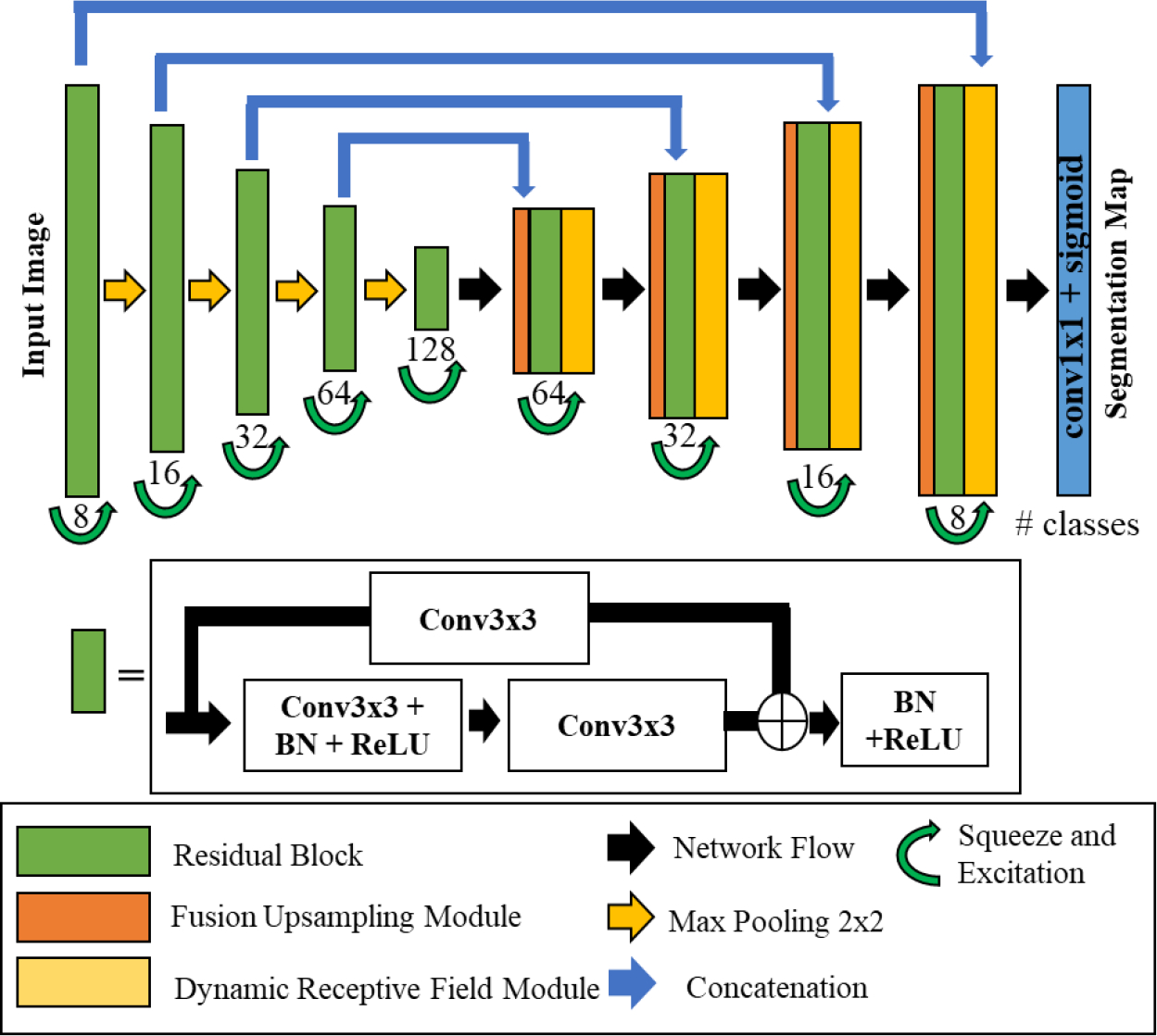
To develop and validate a novel convolutional neural network (CNN) termed "Super U-Net" for medical image segmentation.
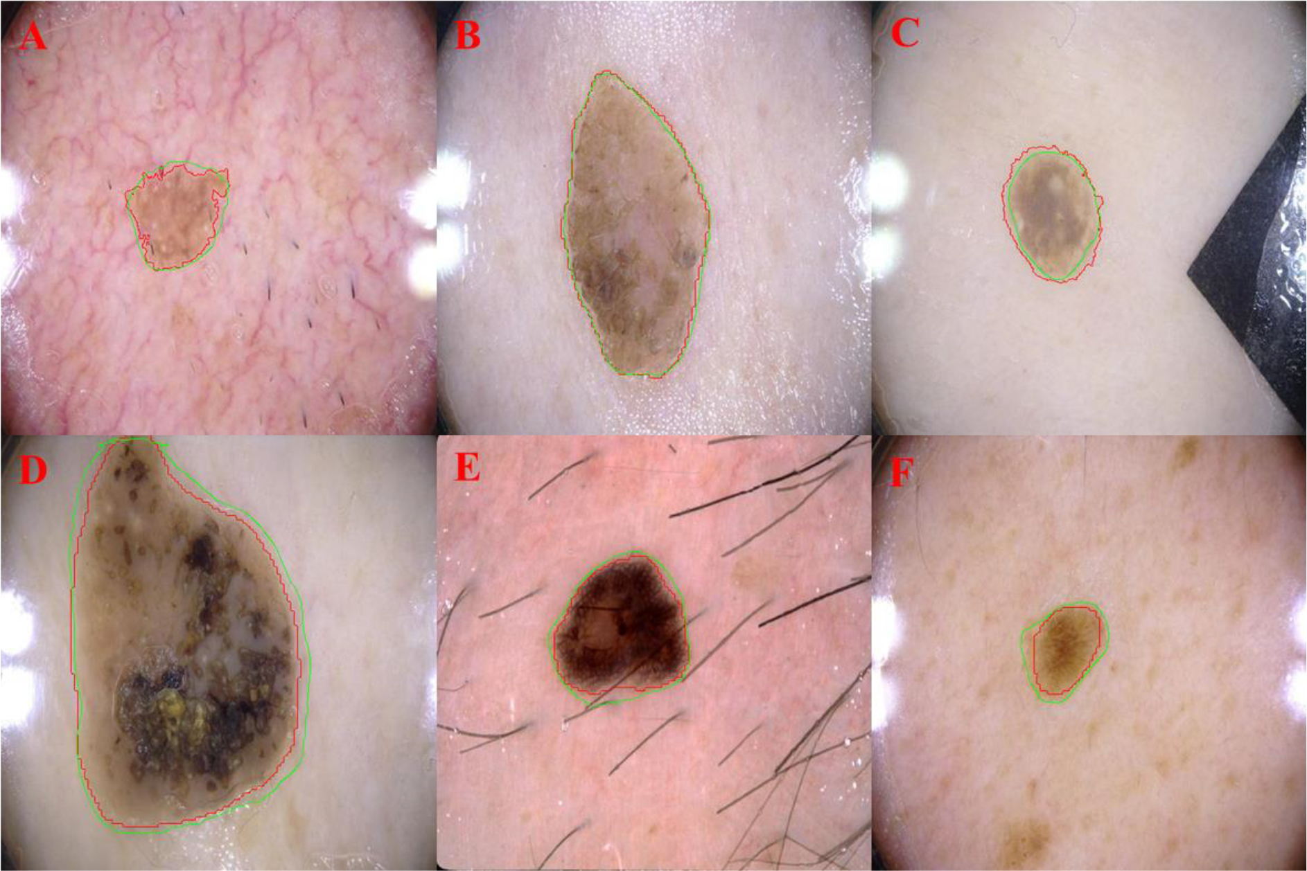
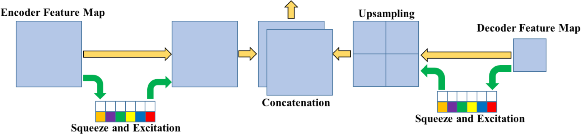
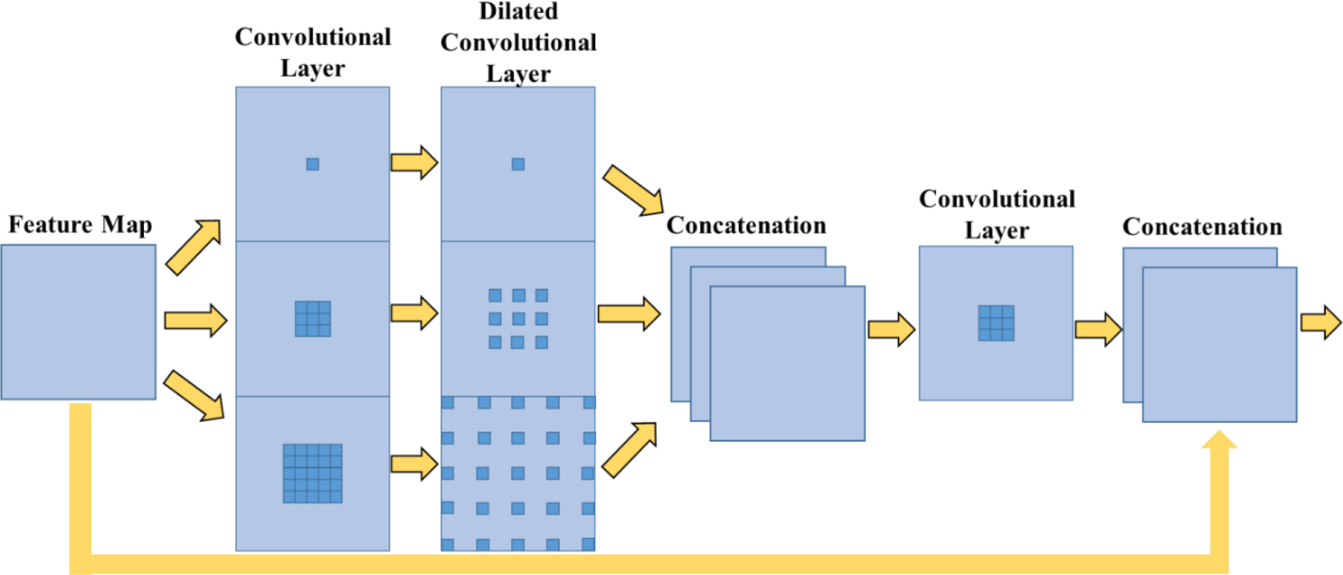
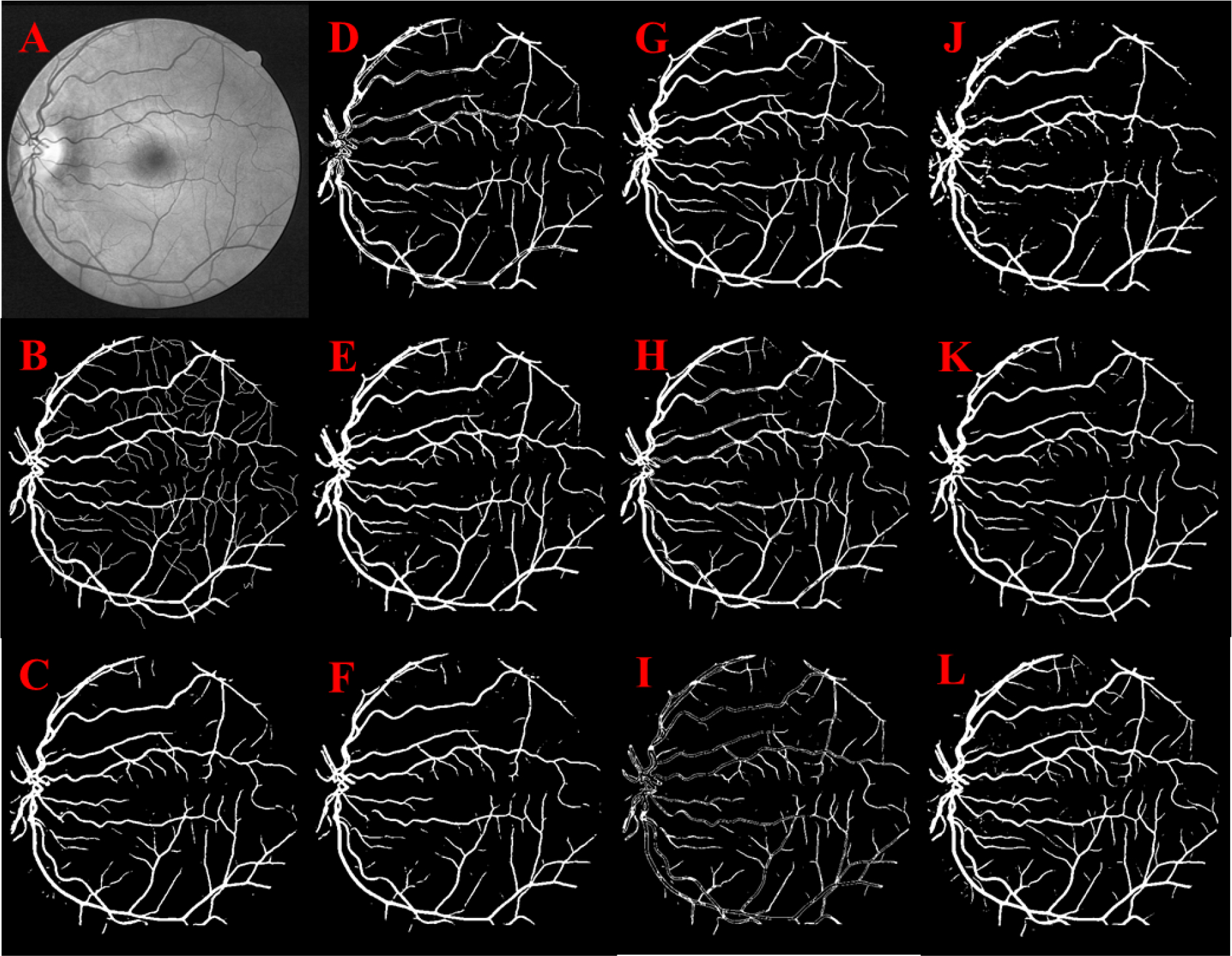
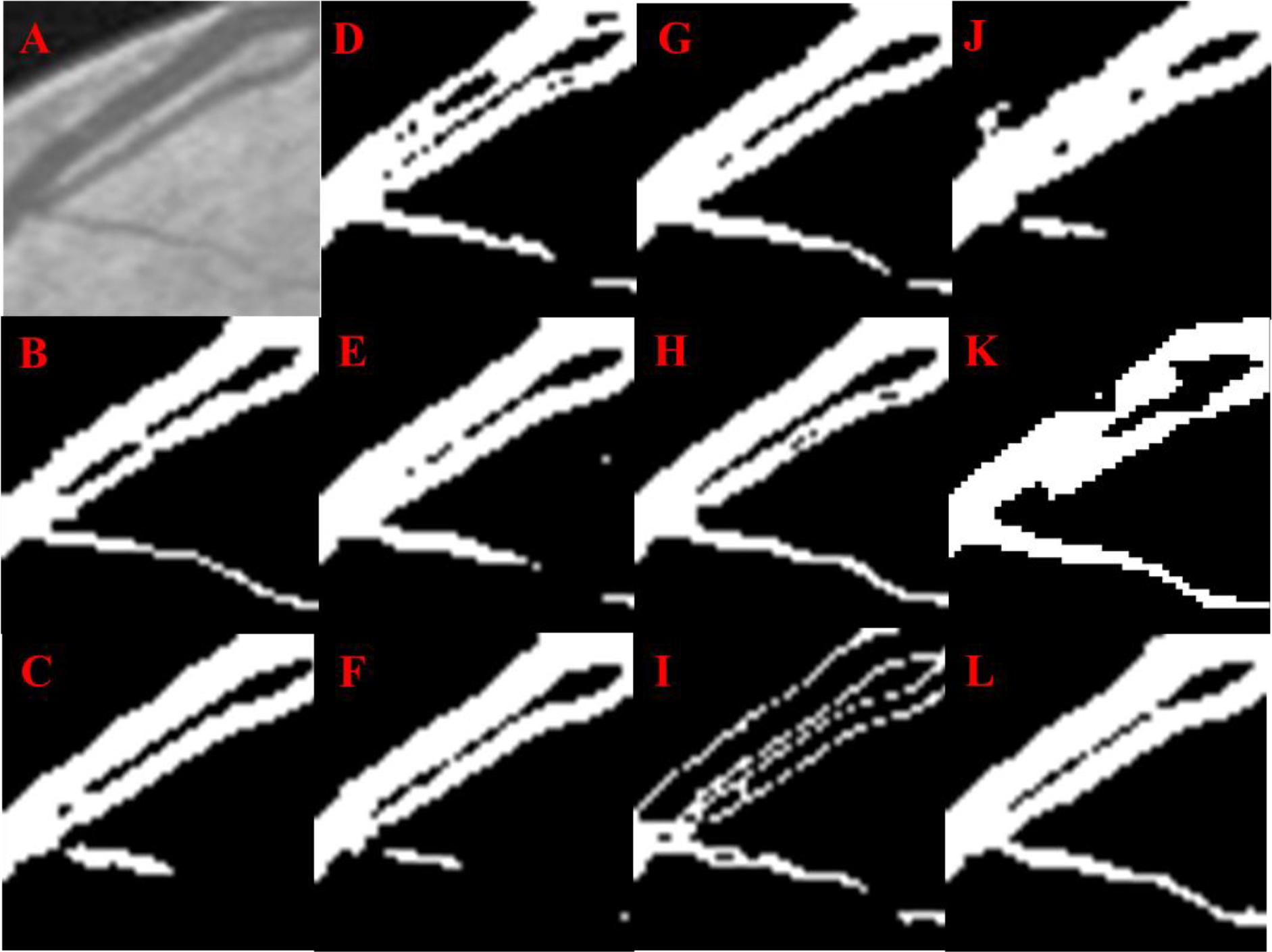
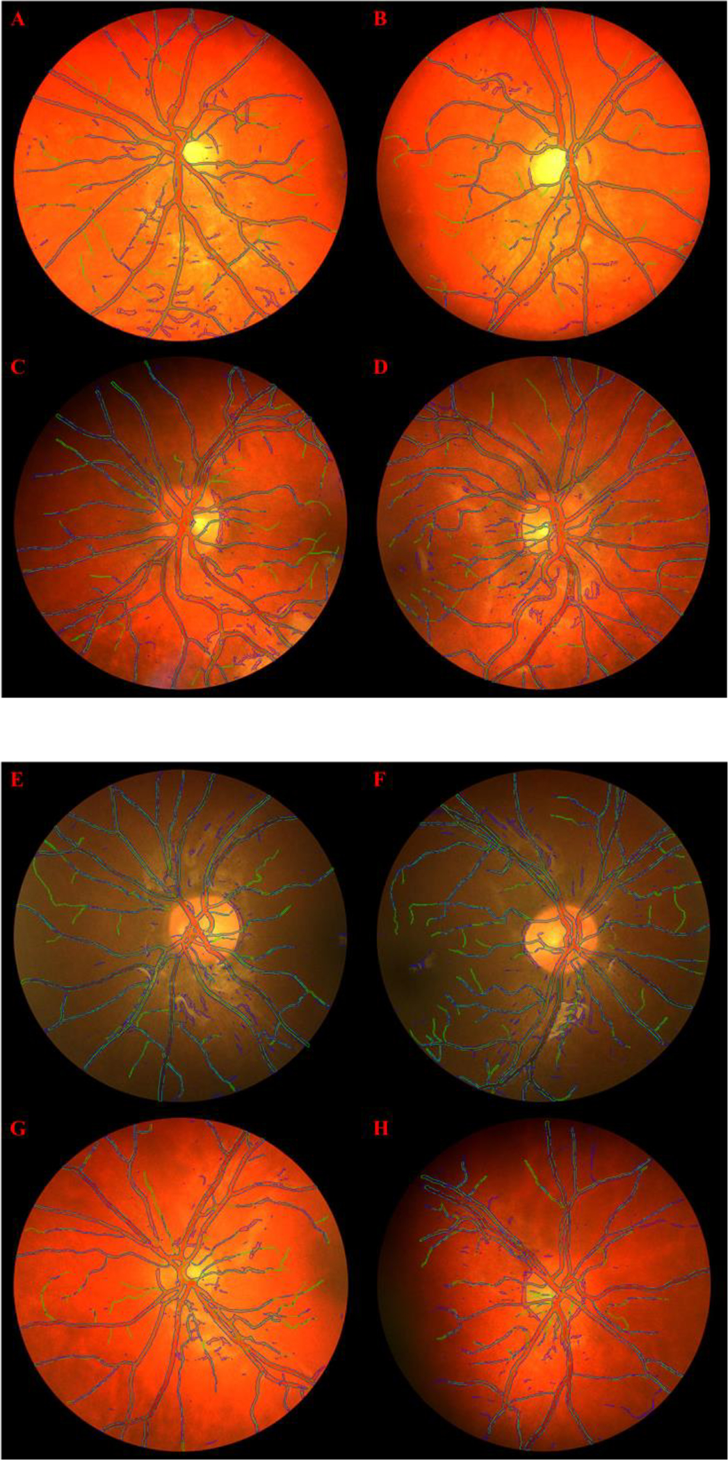
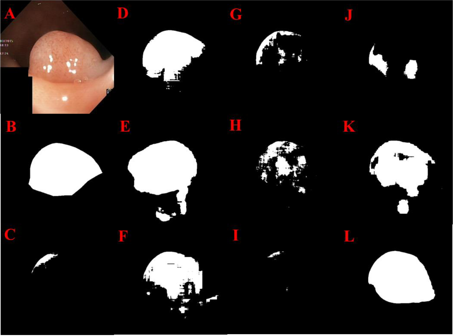
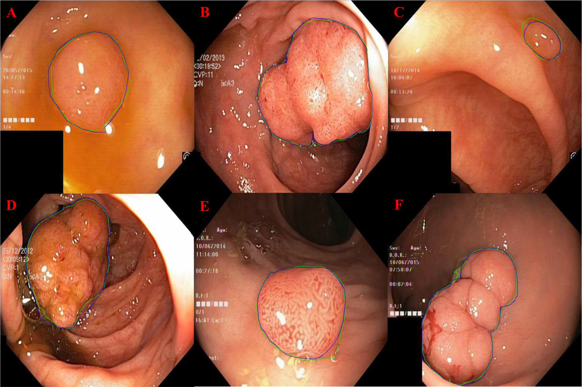
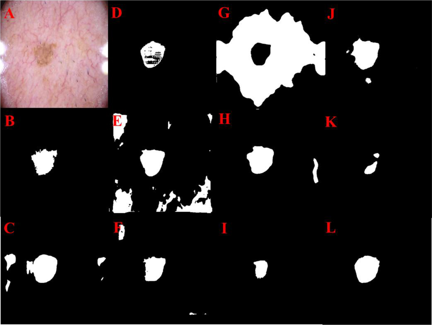
Super U-Net integrates a dynamic receptive field module and a fusion upsampling module into the classical U-Net architecture. The model was developed and tested to segment retinal vessels, gastrointestinal (GI) polyps, skin lesions on several image types (i.e., fundus images, endoscopic images, dermoscopic images). We also trained and tested the traditional U-Net architecture, seven U-Net variants, and two non-U-Net segmentation architectures. K-fold cross-validation was used to evaluate performance. The performance metrics included Dice similarity coefficient (DSC), accuracy, positive predictive value (PPV), and sensitivity.
Super U-Net achieved average DSCs of 0.808±0.0210, 0.752±0.019, 0.804±0.239, and 0.877±0.135 for segmenting retinal vessels, pediatric retinal vessels, GI polyps, and skin lesions, respectively. The Super U-net consistently outperformed U-Net, seven U-Net variants, and two non-U-Net segmentation architectures (p < 0.05).
Dynamic receptive fields and fusion upsampling can significantly improve image segmentation performance.
开发并验证一种名为“超级U-Net”的新型卷积神经网络(CNN)用于医学图像分割。
超级U-Net将动态感受野模块和融合上采样模块集成到经典的U-Net架构中。该模型被开发并测试用于分割视网膜血管、胃肠道(GI)息肉、几种图像类型(即眼底图像、内镜图像、皮肤镜图像)上的皮肤病变。我们还训练并测试了传统的U-Net架构、七种U-Net变体以及两种非U-Net分割架构。采用K折交叉验证来评估性能。性能指标包括Dice相似系数(DSC)、准确率、阳性预测值(PPV)和灵敏度。
超级U-Net在分割视网膜血管、儿童视网膜血管、GI息肉和皮肤病变时,平均DSC分别达到0.808±0.0210、0.752±0.019、0.804±0.239和0.877±0.135。超级U-Net始终优于U-Net、七种U-Net变体以及两种非U-Net分割架构(p<0.05)。
动态感受野和融合上采样可显著提高图像分割性能。