Li Yongfeng, Liu Huawei, Wang Chao, Yan Rongzeng, Xiang Lei, Mu Xiaodan, Zheng Lingling, Liu Changkui, Hu Min
Department of Stomatology, The First Medical Center of PLA General Hospital, Beijing, China.
Beijing Advanced Innovation Center for Biomedical Engineering, Beihang University, Beijing, 100083, China.
NPJ Regen Med. 2023 Jul 24;8(1):38. doi: 10.1038/s41536-023-00308-0.
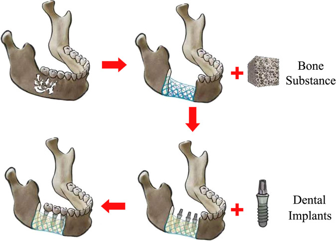
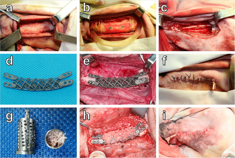
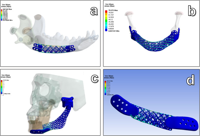
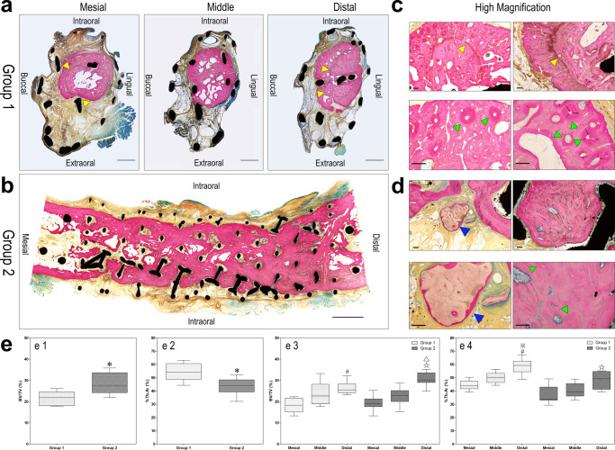
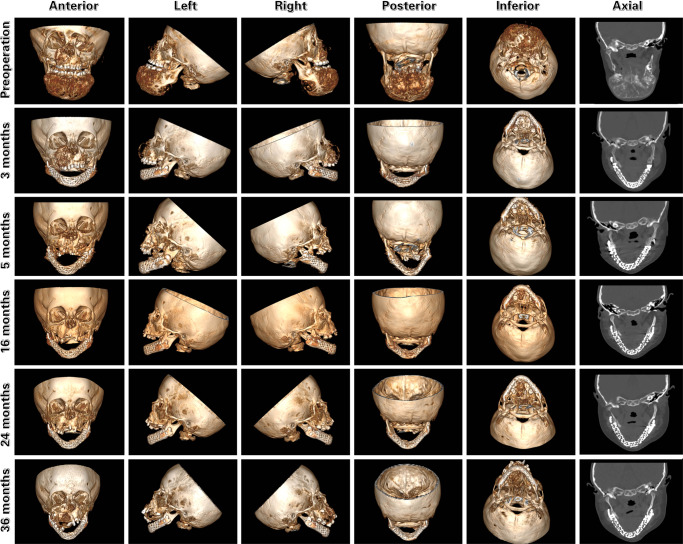
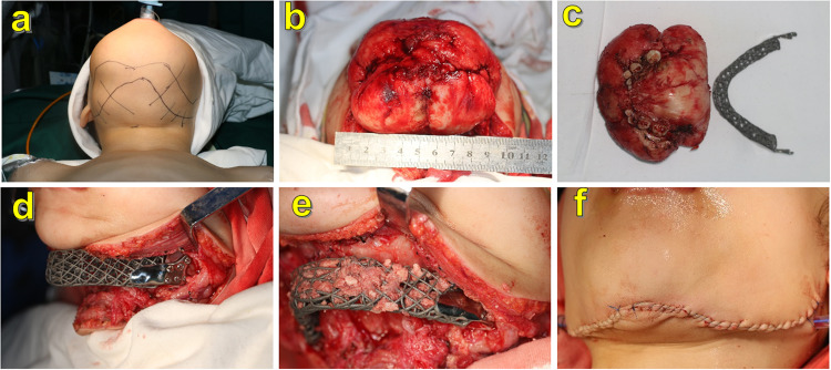
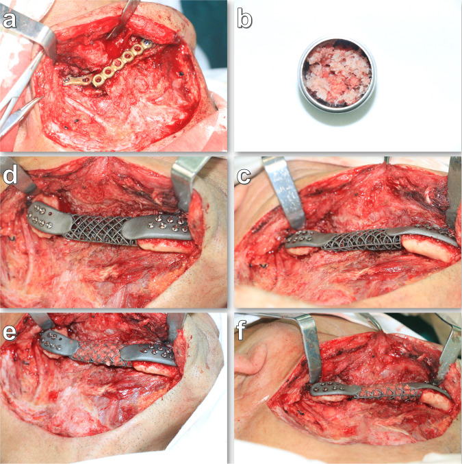
Bone fusion of defect broken ends is the basis of the functional reconstruction of critical maxillofacial segmental bone defects. However, the currently available treatments do not easily achieve this goal. Therefore, this study aimed to fabricate 3D-printing titanium grid scaffolds, which possess sufficient pores and basic biomechanical strength to facilitate osteogenesis in order to accomplish bone fusion in mandibular segmental bone defects. The clinical trial was approved and supervised by the Medical Ethics Committee of the Chinese PLA General Hospital on March 28th, 2019 (Beijing, China. approval No. S2019-065-01), and registered in the clinical trials registry platform (registration number: ChiCTR2300072209). Titanium grid scaffolds were manufactured using selective laser melting and implanted in 20 beagle dogs with mandibular segmental defects. Half of the animals were treated with autologous bone chips and bone substances incorporated into the scaffolds; no additional filling was used for the rest of the animals. After 18 months of observation, radiological scanning and histological analysis in canine models revealed that the pores of regenerated bone were filled with titanium grid scaffolds and bone broken ends were integrated. Furthermore, three patients were treated with similar titanium grid scaffold implants in mandibular segmental defects; no mechanical complications were observed, and similar bone regeneration was observed in the reconstructed patients' mandibles in the clinic. These results demonstrated that 3D-printing titanium grid scaffolds with sufficient pores and basic biomechanical strength could facilitate bone regeneration in large-segment mandibular bone defects.
缺损断端的骨融合是颌面关键节段性骨缺损功能重建的基础。然而,目前可用的治疗方法不易实现这一目标。因此,本研究旨在制造具有足够孔隙率和基本生物力学强度以促进成骨的3D打印钛网支架,从而实现下颌节段性骨缺损的骨融合。该临床试验于2019年3月28日获得中国人民解放军总医院医学伦理委员会的批准和监督(中国北京。批准号:S2019 - 065 - 01),并在临床试验注册平台注册(注册号:ChiCTR2300072209)。使用选择性激光熔化制造钛网支架,并植入20只患有下颌节段性缺损的比格犬体内。一半的动物接受自体骨屑和掺入支架中的骨物质治疗;其余动物未进行额外填充。经过18个月的观察,犬模型的放射学扫描和组织学分析显示,再生骨的孔隙被钛网支架填充,骨断端融合。此外,3例下颌节段性缺损患者接受了类似的钛网支架植入治疗;未观察到机械并发症,且临床观察到重建患者下颌骨中有类似的骨再生。这些结果表明,具有足够孔隙率和基本生物力学强度的3D打印钛网支架可促进大节段下颌骨缺损的骨再生。