• 文献检索
  • 文档翻译
  • 深度研究
  • 学术资讯
  • Suppr Zotero 插件Zotero 插件
  • 邀请有礼
  • 套餐&价格
  • 历史记录
应用&插件
Suppr Zotero 插件Zotero 插件浏览器插件Mac 客户端Windows 客户端微信小程序
定价
高级版会员购买积分包购买API积分包
服务
文献检索文档翻译深度研究API 文档MCP 服务
关于我们
关于 Suppr公司介绍联系我们用户协议隐私条款
关注我们

Suppr 超能文献

核心技术专利:CN118964589B侵权必究
粤ICP备2023148730 号-1Suppr @ 2026

文献检索

告别复杂PubMed语法,用中文像聊天一样搜索,搜遍4000万医学文献。AI智能推荐,让科研检索更轻松。

立即免费搜索

文件翻译

保留排版,准确专业,支持PDF/Word/PPT等文件格式,支持 12+语言互译。

免费翻译文档

深度研究

AI帮你快速写综述,25分钟生成高质量综述,智能提取关键信息,辅助科研写作。

立即免费体验

定义棕色脂肪组织衰老诱导的线粒体嵴形态变化。

Defining Mitochondrial Cristae Morphology Changes Induced by Aging in Brown Adipose Tissue.

机构信息

Department of Molecular Physiology and Biophysics, Vanderbilt University, Nashville, TN, 37232, USA.

Department of Internal Medicine, University of Iowa, Iowa City, IA, 52242, USA.

出版信息

Adv Biol (Weinh). 2024 Jan;8(1):e2300186. doi: 10.1002/adbi.202300186. Epub 2023 Aug 21.

DOI:10.1002/adbi.202300186
PMID:37607124
原文链接:https://pmc.ncbi.nlm.nih.gov/articles/PMC10869235/
Abstract

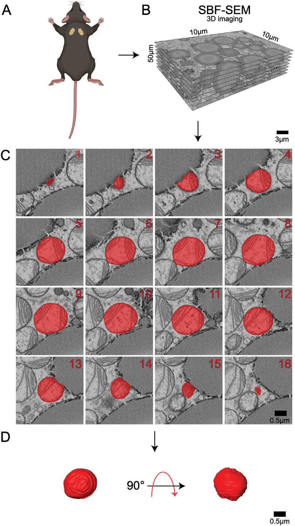
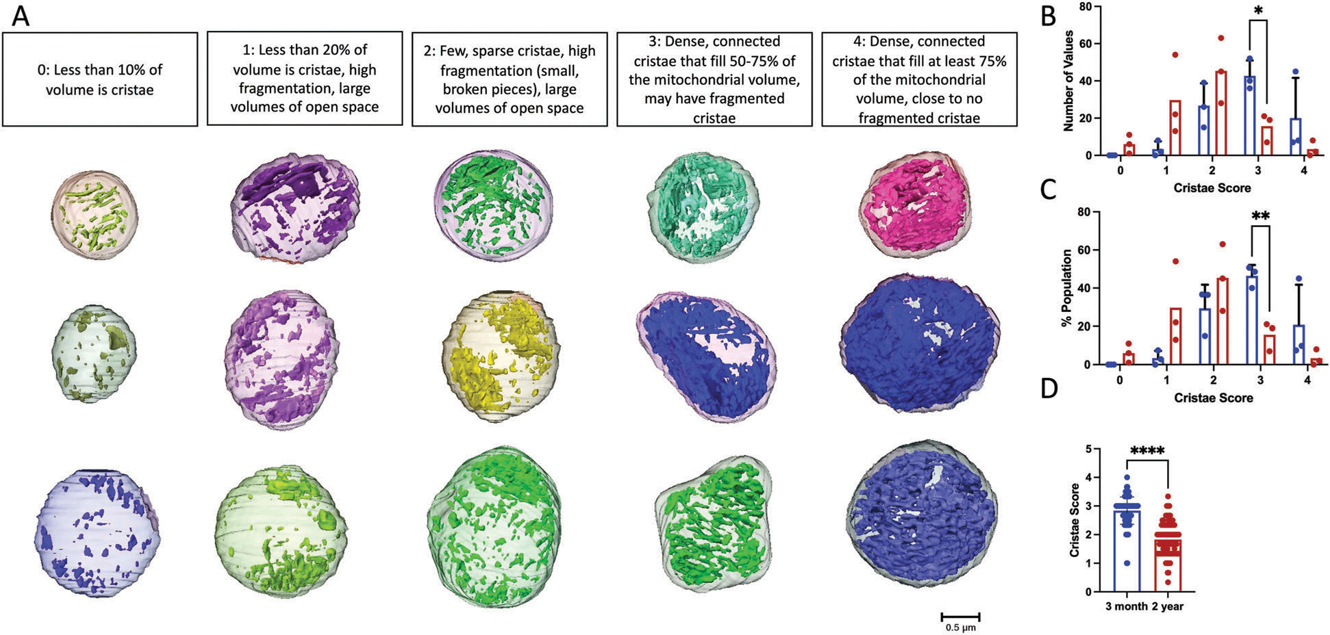
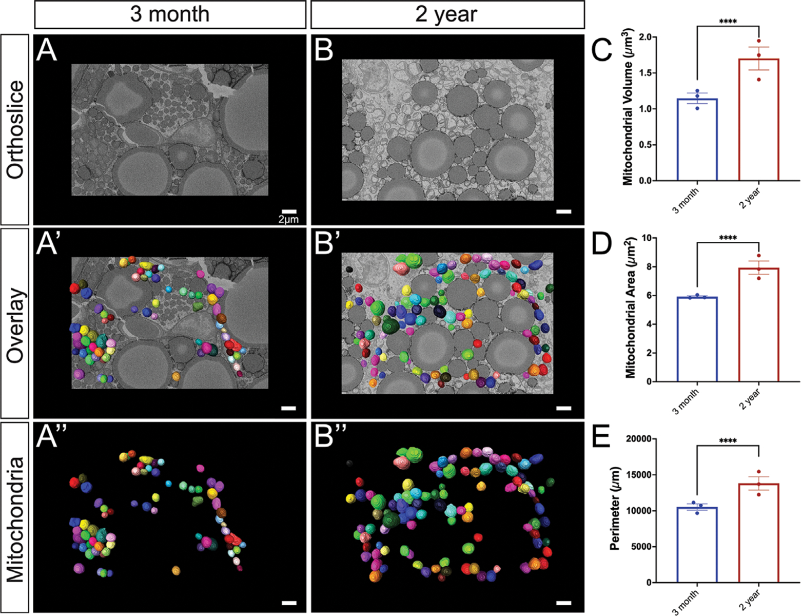
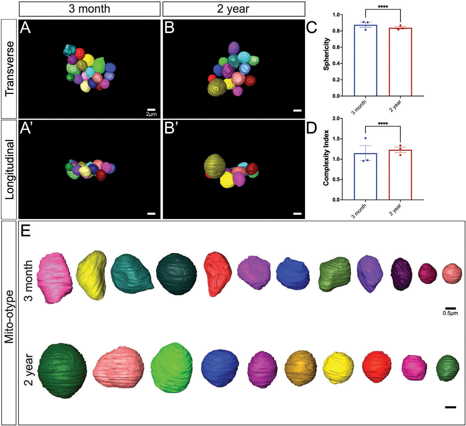
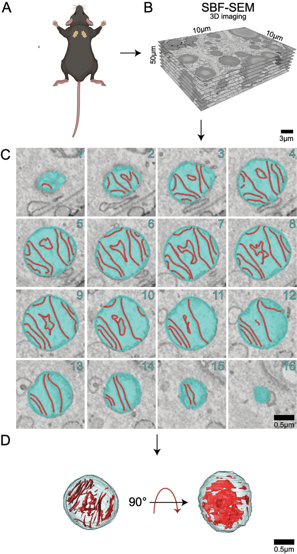
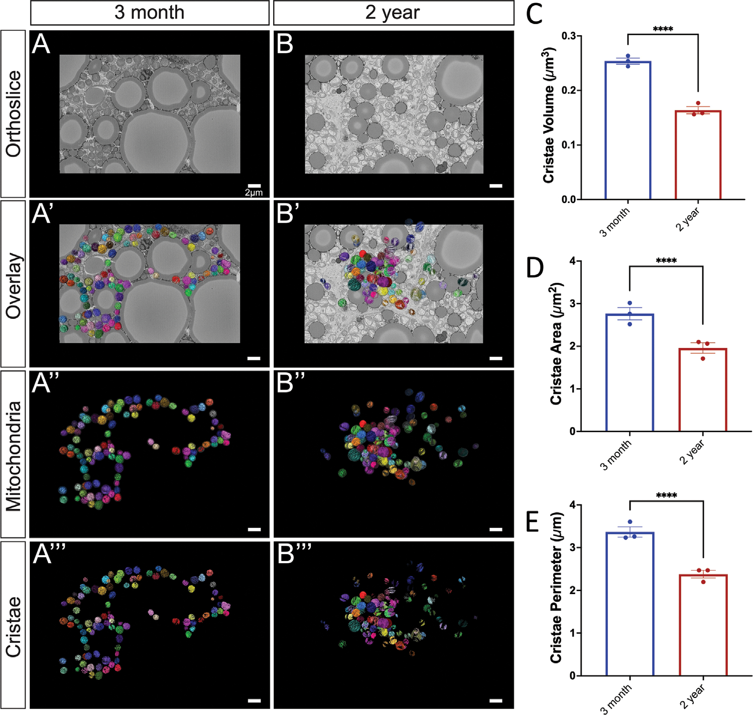
Mitochondria are required for energy production and even give brown adipose tissue (BAT) its characteristic color due to their high iron content and abundance. The physiological function and bioenergetic capacity of mitochondria are connected to the structure, folding, and organization of its inner-membrane cristae. During the aging process, mitochondrial dysfunction is observed, and the regulatory balance of mitochondrial dynamics is often disrupted, leading to increased mitochondrial fragmentation in aging cells. Therefore, it is hypothesized that significant morphological changes in BAT mitochondria and cristae will be present with aging. A quantitative 3D electron microscopy approach is developed to map cristae network organization in mouse BAT to test this hypothesis. Using this methodology, the 3D morphology of mitochondrial cristae is investigated in adult (3-month) and aged (2-year) murine BAT tissue via serial block face-scanning electron microscopy (SBF-SEM) and 3D reconstruction software for manual segmentation, analysis, and quantification. Upon investigation, an increase is found in mitochondrial volume, surface area, and complexity and decreased sphericity in aged BAT, alongside significant decreases in cristae volume, area, perimeter, and score. Overall, these data define the nature of the mitochondrial structure in murine BAT across aging.

摘要

线粒体是能量产生所必需的,由于其含铁量高且丰富,甚至赋予了棕色脂肪组织(BAT)其特征性的颜色。线粒体的生理功能和生物能量能力与其内膜嵴的结构、折叠和组织有关。在衰老过程中,观察到线粒体功能障碍,线粒体动力学的调节平衡经常被打破,导致衰老细胞中线粒体的碎片化增加。因此,人们假设 BAT 线粒体和嵴的显著形态变化将随着衰老而出现。开发了一种定量的 3D 电子显微镜方法来绘制小鼠 BAT 中的嵴网络组织,以验证这一假设。使用这种方法,通过连续块面扫描电子显微镜(SBF-SEM)和用于手动分割、分析和量化的 3D 重建软件,对成年(3 个月)和老年(2 岁)小鼠 BAT 组织中的线粒体嵴的 3D 形态进行了研究。经过研究,发现老年 BAT 中线粒体体积、表面积和复杂性增加,球形度降低,同时嵴体积、面积、周长和分数显著降低。总的来说,这些数据定义了在衰老过程中,小鼠 BAT 中线粒体结构的性质。

https://cdn.ncbi.nlm.nih.gov/pmc/blobs/c23a/10869235/39a6296577ee/nihms-1962717-f0006.jpg
https://cdn.ncbi.nlm.nih.gov/pmc/blobs/c23a/10869235/622a0fc71437/nihms-1962717-f0001.jpg
https://cdn.ncbi.nlm.nih.gov/pmc/blobs/c23a/10869235/4c7ae05d644e/nihms-1962717-f0002.jpg
https://cdn.ncbi.nlm.nih.gov/pmc/blobs/c23a/10869235/572d578686a9/nihms-1962717-f0003.jpg
https://cdn.ncbi.nlm.nih.gov/pmc/blobs/c23a/10869235/0b4bc25f08af/nihms-1962717-f0004.jpg
https://cdn.ncbi.nlm.nih.gov/pmc/blobs/c23a/10869235/9584168d940f/nihms-1962717-f0005.jpg
https://cdn.ncbi.nlm.nih.gov/pmc/blobs/c23a/10869235/39a6296577ee/nihms-1962717-f0006.jpg

相似文献

1
Defining Mitochondrial Cristae Morphology Changes Induced by Aging in Brown Adipose Tissue.定义棕色脂肪组织衰老诱导的线粒体嵴形态变化。
Adv Biol (Weinh). 2024 Jan;8(1):e2300186. doi: 10.1002/adbi.202300186. Epub 2023 Aug 21.
2
Defining Mitochondrial Cristae Morphology Changes Induced by Aging in Brown Adipose Tissue.定义棕色脂肪组织衰老诱导的线粒体嵴形态变化。
bioRxiv. 2023 Aug 2:2023.05.12.540609. doi: 10.1101/2023.05.12.540609.
3
Quantitative assessment of morphological changes in lipid droplets and lipid-mito interactions with aging in brown adipose.定量评估棕色脂肪中脂滴形态变化和脂-线粒体相互作用随年龄的变化。
J Cell Physiol. 2024 Sep;239(9):e31340. doi: 10.1002/jcp.31340. Epub 2024 Aug 13.
4
Quantitative Assessment of Morphological Changes in Lipid Droplets and Lipid-Mito Interactions with Aging in Brown Adipose.棕色脂肪中脂滴形态变化及脂质-线粒体相互作用随衰老的定量评估
bioRxiv. 2024 May 21:2023.09.23.559135. doi: 10.1101/2023.09.23.559135.
5
3D reconstruction of murine mitochondria reveals changes in structure during aging linked to the MICOS complex.鼠线粒体的 3D 重建揭示了与 MICOS 复合物相关的衰老过程中结构的变化。
Aging Cell. 2023 Dec;22(12):e14009. doi: 10.1111/acel.14009. Epub 2023 Nov 13.
6
Jmjd maintains the morphology and function of lipid droplets and mitochondria of brown adipocytes in mice.Jmjd维持小鼠棕色脂肪细胞中脂滴和线粒体的形态及功能。
Zhong Nan Da Xue Xue Bao Yi Xue Ban. 2020 Nov 28;45(11):1298-1307. doi: 10.11817/j.issn.1672-7347.2020.190364.
7
Lipid droplet remodeling and interaction with mitochondria in mouse brown adipose tissue during cold treatment.冷处理期间小鼠棕色脂肪组织中脂滴重塑及其与线粒体的相互作用
Biochim Biophys Acta. 2015 May;1853(5):918-28. doi: 10.1016/j.bbamcr.2015.01.020. Epub 2015 Feb 2.
8
A semi-automatic method for extracting mitochondrial cristae characteristics from 3D focused ion beam scanning electron microscopy data.一种从 3D 聚焦离子束扫描电子显微镜数据中提取线粒体嵴特征的半自动方法。
Commun Biol. 2024 Mar 28;7(1):377. doi: 10.1038/s42003-024-06045-4.
9
Brown adipose tissue development and metabolism in ruminants.反刍动物的棕色脂肪组织发育与代谢
J Anim Sci. 2004 Mar;82(3):942-54. doi: 10.2527/2004.823942x.
10
A lack of ChREBP inhibits mitochondrial cristae formation in brown adipose tissue.ChREBP 的缺乏会抑制棕色脂肪组织中线粒体嵴的形成。
Mol Cell Biochem. 2021 Oct;476(10):3577-3590. doi: 10.1007/s11010-021-04178-2. Epub 2021 May 21.

引用本文的文献

1
3D Mitochondrial Structure in Aging Human Skeletal Muscle: Insights Into MFN-2-Mediated Changes.衰老人类骨骼肌中的三维线粒体结构:对MFN-2介导变化的见解
Aging Cell. 2025 Apr 25:e70054. doi: 10.1111/acel.70054.
2
The ultrastructural and proteomic analysis of mitochondria-associated endoplasmic reticulum membrane in the midbrain of a Parkinson's disease mouse model.帕金森病小鼠模型中脑线粒体相关内质网膜的超微结构和蛋白质组学分析
Aging Cell. 2025 Apr;24(4):e14436. doi: 10.1111/acel.14436. Epub 2024 Nov 29.
3
A workshop to enrich physiological understanding through hands-on learning about mitochondria-endoplasmic reticulum contact sites.

本文引用的文献

1
Correction: Garza-Lopez et al. Protocols for Generating Surfaces and Measuring 3D Organelle Morphology Using Amira. 2022, , 65.更正:加尔萨-洛佩斯等人。使用Amira生成表面和测量三维细胞器形态的方案。2022年,第65页。
Cells. 2023 May 10;12(10):1356. doi: 10.3390/cells12101356.
2
Serial Block Face-Scanning Electron Microscopy as a Burgeoning Technology.序列块面扫描电子显微镜技术的兴起。
Adv Biol (Weinh). 2023 Aug;7(8):e2300139. doi: 10.1002/adbi.202300139. Epub 2023 May 28.
3
A Comprehensive Approach to Sample Preparation for Electron Microscopy and the Assessment of Mitochondrial Morphology in Tissue and Cultured Cells.
通过动手学习线粒体-内质网接触点来丰富生理学理解的研讨会。
Adv Physiol Educ. 2024 Dec 1;48(4):808-817. doi: 10.1152/advan.00271.2023. Epub 2024 Sep 5.
4
Mitochondrial heterogeneity and crosstalk in aging: Time for a paradigm shift?线粒体异质性与衰老中的相互作用:是否需要范式转变?
Aging Cell. 2024 Oct;23(10):e14296. doi: 10.1111/acel.14296. Epub 2024 Aug 26.
5
Mitochondrial Structure and Function in Human Heart Failure.线粒体结构和功能在人类心力衰竭中的作用。
Circ Res. 2024 Jul 5;135(2):372-396. doi: 10.1161/CIRCRESAHA.124.323800. Epub 2024 Jul 4.
6
The MICOS Complex Regulates Mitochondrial Structure and Oxidative Stress During Age-Dependent Structural Deficits in the Kidney.MICOS复合体在肾脏年龄依赖性结构缺陷期间调节线粒体结构和氧化应激。
bioRxiv. 2024 Jun 12:2024.06.09.598108. doi: 10.1101/2024.06.09.598108.
7
Connection Between HIV and Mitochondria in Cardiovascular Disease and Implications for Treatments.HIV 与心血管疾病中线粒体的关联及其治疗意义。
Circ Res. 2024 May 24;134(11):1581-1606. doi: 10.1161/CIRCRESAHA.124.324296. Epub 2024 May 23.
8
New Mediators in the Crosstalk between Different Adipose Tissues.不同脂肪组织间串扰的新介质。
Int J Mol Sci. 2024 Apr 25;25(9):4659. doi: 10.3390/ijms25094659.
9
ATF4-dependent increase in mitochondrial-endoplasmic reticulum tethering following OPA1 deletion in skeletal muscle.OPA1 缺失后骨骼肌中线粒体-内质网连接增加依赖于 ATF4。
J Cell Physiol. 2024 Apr;239(4):e31204. doi: 10.1002/jcp.31204. Epub 2024 Feb 28.
10
Mitochondria in disease: changes in shapes and dynamics.线粒体在疾病中的作用:形态和动力学的变化。
Trends Biochem Sci. 2024 Apr;49(4):346-360. doi: 10.1016/j.tibs.2024.01.011. Epub 2024 Feb 23.
一种全面的电子显微镜样品制备方法及组织和培养细胞中线粒体形态评估。
Adv Biol (Weinh). 2023 Oct;7(10):e2200202. doi: 10.1002/adbi.202200202. Epub 2023 May 4.
4
Systematic Transmission Electron Microscopy-Based Identification and 3D Reconstruction of Cellular Degradation Machinery.基于系统透射电子显微镜的细胞降解机制的鉴定和 3D 重构。
Adv Biol (Weinh). 2023 Jun;7(6):e2200221. doi: 10.1002/adbi.202200221. Epub 2023 Mar 3.
5
How does density of the inner mitochondrial membrane influence mitochondrial performance?内线粒体膜的密度如何影响线粒体的性能?
Am J Physiol Regul Integr Comp Physiol. 2023 Feb 1;324(2):R242-R248. doi: 10.1152/ajpregu.00254.2022. Epub 2022 Dec 26.
6
Brown Adipose Tissue-A Translational Perspective.棕色脂肪组织——转化视角。
Endocr Rev. 2023 Mar 4;44(2):143-192. doi: 10.1210/endrev/bnac015.
7
Therapeutic Strategies Targeting Mitochondrial Calcium Signaling: A New Hope for Neurological Diseases?针对线粒体钙信号的治疗策略:神经系统疾病的新希望?
Antioxidants (Basel). 2022 Jan 15;11(1):165. doi: 10.3390/antiox11010165.
8
Spermidine supplementation influences mitochondrial number and morphology in the heart of aged mice.亚精胺补充影响衰老小鼠心脏中线粒体的数量和形态。
J Anat. 2023 Jan;242(1):91-101. doi: 10.1111/joa.13618. Epub 2021 Dec 27.
9
Mitochondrial dysfunction plays an essential role in remodeling aging adipose tissue.线粒体功能障碍在重塑衰老脂肪组织中起着至关重要的作用。
Mech Ageing Dev. 2021 Dec;200:111598. doi: 10.1016/j.mad.2021.111598. Epub 2021 Nov 9.
10
A Universal Approach to Analyzing Transmission Electron Microscopy with ImageJ.利用 ImageJ 进行透射电子显微镜分析的通用方法
Cells. 2021 Aug 24;10(9):2177. doi: 10.3390/cells10092177.