Ancuta Diana Larisa, Alexandru Diana Mihaela, Crivineanu Maria, Coman Cristin
Faculty of Veterinary Medicine, University of Agronomic Sciences and Veterinary Medicine, 050097 Bucharest, Romania.
Cantacuzino National Medical Military Institute for Research and Development, 050096 Bucharest, Romania.
Biomedicines. 2023 Jul 25;11(8):2098. doi: 10.3390/biomedicines11082098.
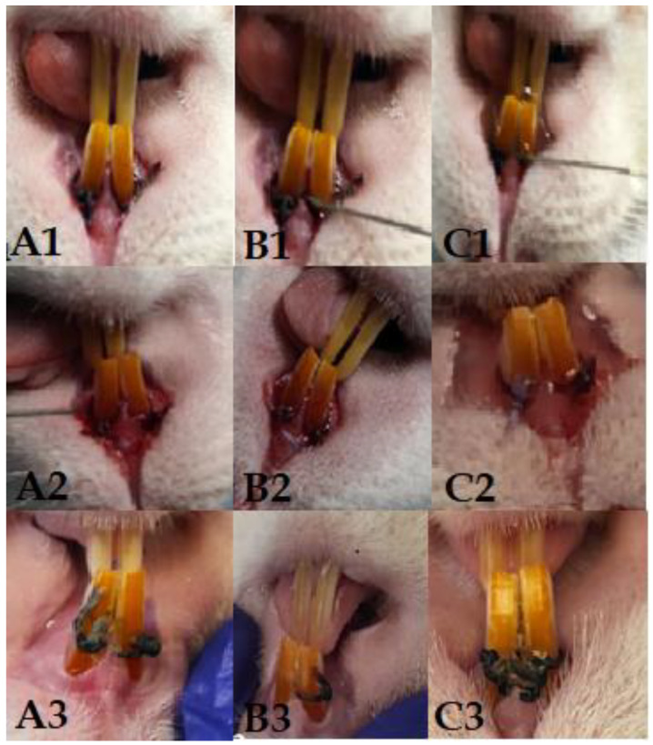
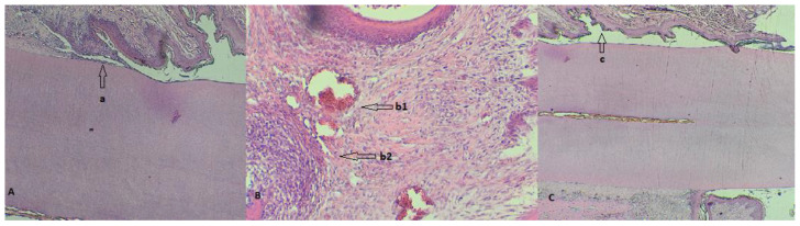
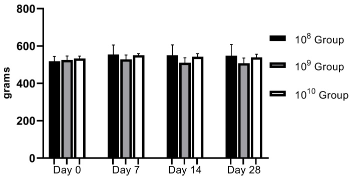
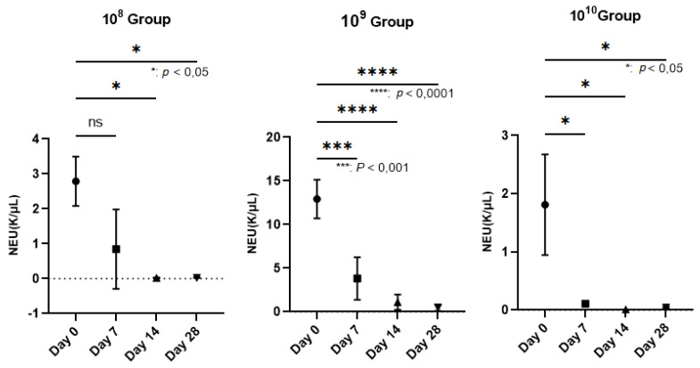
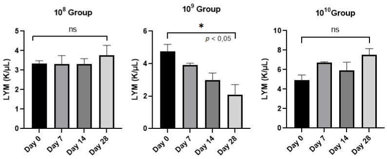
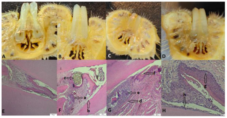
Periodontal disease is that condition resulting in the destruction of periodontal tissues, bone resorption, and tooth loss, the etiology of which is linked to immunological and microbiological factors. The aim of this study was to evaluate the potential trigger of periodontal disease in a rat model using bacterial species incriminated in the pathology of human periodontitis and to establish their optimal concentrations capable of reproducing the disease, with the idea of subsequently developing innovative treatments for the condition. In this study, we included 15 male Wistar rats, aged 20 weeks, which we divided into three groups. In each group, we applied ligatures with gingival retraction wire on the maxillary incisors. The ligature and the gingival sac were contaminated by oral gavage with a mixture of fresh cultures of (A.a), (F.n) and (S.o) in concentrations of 10, 10, and 10 CFU/mL each for 5 days a week for 4 weeks. During the clinical monitoring period of 28 days, overlapped with the period of oral contamination, we followed the expression of clinical signs specific to periodontitis. We also monitored the evolution of body weight and took weekly samples from the oral cavity for the microbiological identification of the tested bacteria and blood samples for hematological examination. At the end of the study, the animals were euthanized, and the ligated incisors were taken for histopathological analysis. The characteristic symptomatology of periodontal disease was expressed from the first week of the study and was maintained until the end, and we were able to identify the bacteria during each examination. Hematologically, the number of neutrophils decreased dramatically ( < 0.0001) in the case of the 10 group, unlike the other groups, as did the number of lymphocytes. Histopathologically, we identified neutrophilic infiltrate in all groups, as well as the presence of coccobacilli, periodontal tissue hyperplasia, and periodontal lysis. In the 10 group, we also observed pulpal tissue with necrotic bone fragments and pyogranulomatous inflammatory reaction. By corroborating the data, we can conclude that for the development of periodontal disease using A.a, F.n, and S.o, a concentration of 10 or 10 CFU/mL is required, which must necessarily contaminate a ligature thread applied to the level of the rat's dental pack.
牙周病是一种导致牙周组织破坏、骨质吸收和牙齿脱落的病症,其病因与免疫和微生物因素有关。本研究的目的是在大鼠模型中,使用与人类牙周炎病理相关的细菌种类评估牙周病的潜在触发因素,并确定能够引发该疾病的最佳浓度,以便随后开发针对该病症的创新治疗方法。在本研究中,我们纳入了15只20周龄的雄性Wistar大鼠,并将它们分为三组。在每组中,我们用上颌切牙的牙龈退缩线进行结扎。通过口服灌胃,用浓度分别为10、10和10 CFU/mL的牙龈卟啉单胞菌(A.a)、具核梭杆菌(F.n)和伴放线放线杆菌(S.o)的新鲜培养物混合物污染结扎线和牙龈囊,每周5天,持续4周。在与口腔污染期重叠的28天临床监测期内,我们观察了牙周炎特有的临床症状的表现。我们还监测了体重的变化,并每周从口腔取样进行受试细菌的微生物鉴定,以及采集血样进行血液学检查。在研究结束时,对动物实施安乐死,并取出结扎的切牙进行组织病理学分析。牙周病的特征性症状从研究的第一周开始出现,并持续到结束,并且我们在每次检查中都能够鉴定出细菌。血液学方面,与其他组不同,10组的中性粒细胞数量显著减少(<0.0001),淋巴细胞数量也减少。组织病理学上,我们在所有组中都发现了嗜中性粒细胞浸润,以及球杆菌的存在、牙周组织增生和牙周溶解。在10组中,我们还观察到牙髓组织伴有坏死骨碎片和脓性肉芽肿性炎症反应。通过综合数据,我们可以得出结论,使用牙龈卟啉单胞菌、具核梭杆菌和伴放线放线杆菌引发牙周病,需要10或10 CFU/mL的浓度,且必须污染应用于大鼠牙列水平处的结扎线。