Song Wei, Guo Changkui, Zhao Yuting, Wang Ya-Chao, Zhu Siwei, Min Changjun, Yuan Xiaocong
Nanophotonics Research Center, Shenzhen Key Laboratory of Micro-Scale Optical Information Technology, Institute of Microscale Optoelectronics & State Key Laboratory of Radio Frequency Heterogeneous, Shenzhen University, Shenzhen 518060, China.
Depart of Neurosurgery, The First Affiliated Hospital of Shenzhen University, Shenzhen Second People's Hospital, Shenzhen 518060, China.
Photoacoustics. 2023 Jun 28;32:100525. doi: 10.1016/j.pacs.2023.100525. eCollection 2023 Aug.
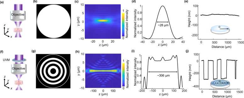
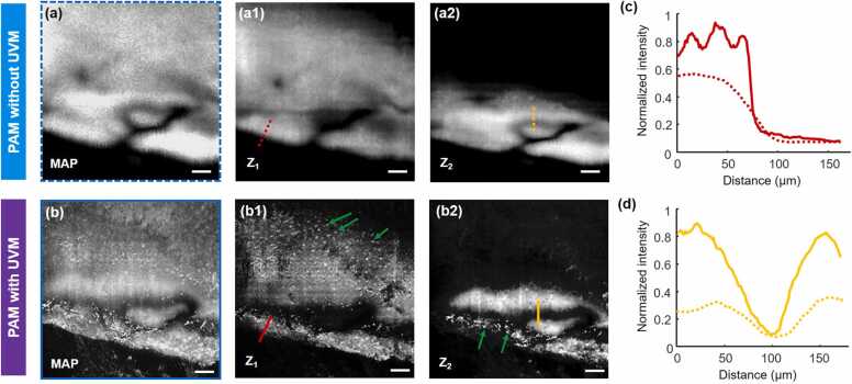
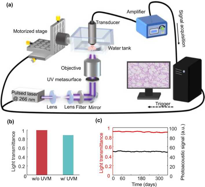
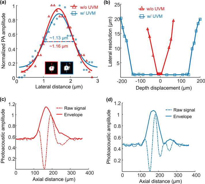
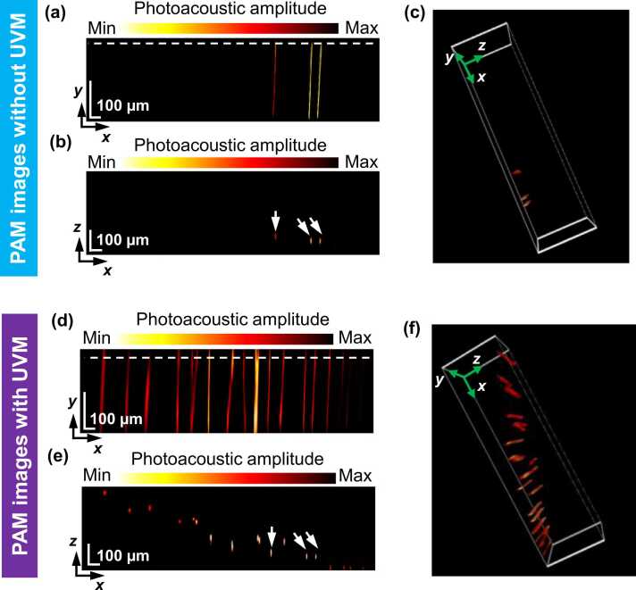
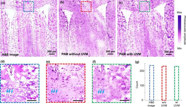
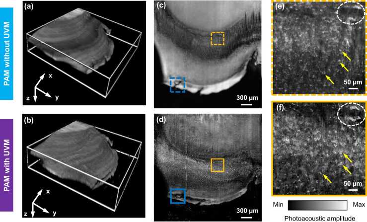
Pathology interpretations of tissue rely on the gold standard of histology imaging, potentially hampering timely access to critical information for diagnosis and management of neoplasms because of tedious sample preparations. Slide-free capture of cell nuclei in unprocessed specimens without staining is preferable; however, inevitable irregular surfaces in fresh tissues results in limitations. An ultraviolet metasurface with the ability to generate an ultraviolet optical focus maintaining < 1.1-µm in lateral resolution and ∼290 µm in depth of field (DOF) is proposed for fast, high resolution, label-free photoacoustic histological imaging of unprocessed tissues with uneven surfaces. Microanatomical characteristics of the cell nuclei can be observed, as demonstrated by the mouse brain samples that were cut by hand and a ∼3 × 3-mm field of view was imaged in ∼27 min. Therefore, ultraviolet metasurface-assisted photoacoustic microscopy is anticipated to benefit intraoperative pathological assessments and basic scientific research by alleviating laborious tissue preparations.
组织的病理学解释依赖于组织学成像的金标准,由于样本制备繁琐,这可能会妨碍及时获取肿瘤诊断和管理的关键信息。在未处理的标本中无染色地无载玻片捕获细胞核是可取的;然而,新鲜组织中不可避免的不规则表面会导致局限性。提出了一种具有产生紫外光焦点能力的紫外超表面,其横向分辨率保持<1.1μm,景深(DOF)约为290μm,用于对表面不平的未处理组织进行快速、高分辨率、无标记的光声组织学成像。可以观察到细胞核的微观解剖特征,如通过手工切割的小鼠脑样本所证明的,在约27分钟内对约3×3毫米的视野进行了成像。因此,预计紫外超表面辅助光声显微镜将通过减轻繁琐的组织制备工作,有益于术中病理评估和基础科学研究。