• 文献检索
  • 文档翻译
  • 深度研究
  • 学术资讯
  • Suppr Zotero 插件Zotero 插件
  • 邀请有礼
  • 套餐&价格
  • 历史记录
应用&插件
Suppr Zotero 插件Zotero 插件浏览器插件Mac 客户端Windows 客户端微信小程序
定价
高级版会员购买积分包购买API积分包
服务
文献检索文档翻译深度研究API 文档MCP 服务
关于我们
关于 Suppr公司介绍联系我们用户协议隐私条款
关注我们

Suppr 超能文献

核心技术专利:CN118964589B侵权必究
粤ICP备2023148730 号-1Suppr @ 2026

文献检索

告别复杂PubMed语法,用中文像聊天一样搜索,搜遍4000万医学文献。AI智能推荐,让科研检索更轻松。

立即免费搜索

文件翻译

保留排版,准确专业,支持PDF/Word/PPT等文件格式,支持 12+语言互译。

免费翻译文档

深度研究

AI帮你快速写综述,25分钟生成高质量综述,智能提取关键信息,辅助科研写作。

立即免费体验

利用机器学习技术对 MRI-ADC 图像的纹理特征进行分析,以区分胶质瘤的级别。

Texture feature analysis of MRI-ADC images to differentiate glioma grades using machine learning techniques.

机构信息

Department of Radiology, Faculty of Medicine, University of Peradeniya, Peradeniya, 20400, Sri Lanka.

Department of Radiography/Radiotherapy, Faculty of Allied Health Sciences, University of Peradeniya, Peradeniya, 20400, Sri Lanka.

出版信息

Sci Rep. 2023 Sep 22;13(1):15772. doi: 10.1038/s41598-023-41353-5.

DOI:10.1038/s41598-023-41353-5
PMID:37737249
原文链接:https://pmc.ncbi.nlm.nih.gov/articles/PMC10517003/
Abstract

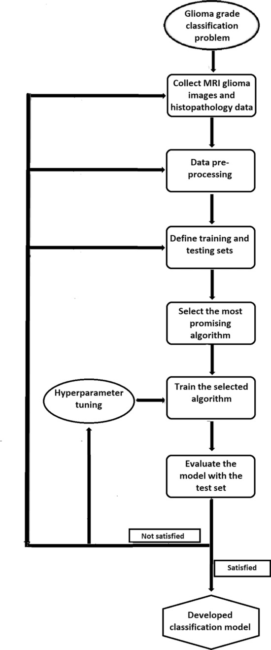
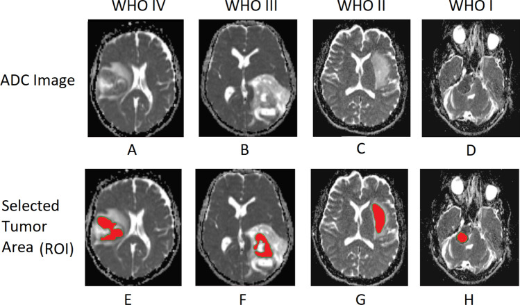
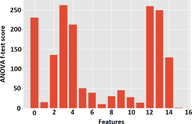
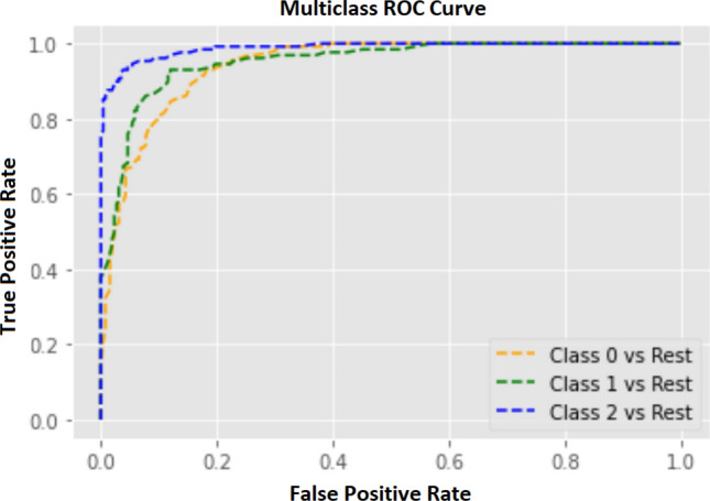
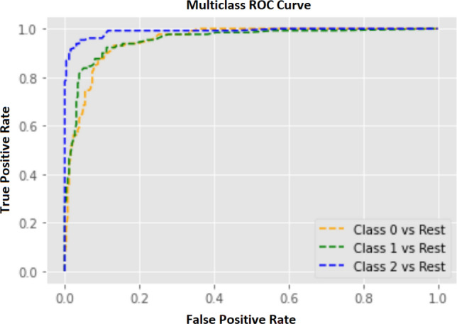
Apparent diffusion coefficient (ADC) of magnetic resonance imaging (MRI) is an indispensable imaging technique in clinical neuroimaging that quantitatively assesses the diffusivity of water molecules within tissues using diffusion-weighted imaging (DWI). This study focuses on developing a robust machine learning (ML) model to predict the aggressiveness of gliomas according to World Health Organization (WHO) grading by analyzing patients' demographics, higher-order moments, and grey level co-occurrence matrix (GLCM) texture features of ADC. A population of 722 labeled MRI-ADC brain image slices from 88 human subjects was selected, where gliomas are labeled as glioblastoma multiforme (WHO-IV), high-grade glioma (WHO-III), and low-grade glioma (WHO I-II). Images were acquired using 3T-MR systems and a region of interest (ROI) was delineated manually over tumor areas. Skewness, kurtosis, and statistical texture features of GLCM (mean, variance, energy, entropy, contrast, homogeneity, correlation, prominence, and shade) were calculated using ADC values within ROI. The ANOVA f-test was utilized to select the best features to train an ML model. The data set was split into training (70%) and testing (30%) sets. The train set was fed into several ML algorithms and selected most promising ML algorithm using K-fold cross-validation. The hyper-parameters of the selected algorithm were optimized using random grid search technique. Finally, the performance of the developed model was assessed by calculating accuracy, precision, recall, and F1 values reported for the test set. According to the ANOVA f-test, three attributes; patient gender (1.48), GLCM energy (9.48), and correlation (13.86) that performed minimum scores were excluded from the dataset. Among the tested algorithms, the random forest classifier(0.8772 ± 0.0237) performed the highest mean-cross-validation score and selected to build the ML model which was able to predict tumor categories with an accuracy of 88.14% over the test set. The study concludes that the developed ML model using the above features except for patient gender, GLCM energy, and correlation, has high prediction accuracy in glioma grading. Therefore, the outcomes of this study enable to development of advanced tumor classification applications that assist in the decision-making process in a real-time clinical environment.

摘要

磁共振成像(MRI)的表观扩散系数(ADC)是临床神经影像学中不可或缺的成像技术,它使用扩散加权成像(DWI)定量评估组织内水分子的扩散性。本研究专注于开发一个稳健的机器学习(ML)模型,通过分析患者的人口统计学、高阶矩和灰度共生矩阵(GLCM)纹理特征的 ADC,根据世界卫生组织(WHO)分级预测胶质瘤的侵袭性。从 88 名人类受试者中选择了 722 个标记的 MRI-ADC 脑图像切片的人群,其中胶质瘤被标记为多形性胶质母细胞瘤(WHO-IV)、高级别胶质瘤(WHO-III)和低级别胶质瘤(WHO I-II)。图像是使用 3T-MR 系统采集的,手动在肿瘤区域划定感兴趣区域(ROI)。使用 ROI 内的 ADC 值计算偏度、峰度和 GLCM 的统计纹理特征(均值、方差、能量、熵、对比度、同质性、相关性、显著性和阴影)。使用方差分析(ANOVA)f 检验选择最佳特征来训练 ML 模型。数据集分为训练集(70%)和测试集(30%)。将训练集输入到几种 ML 算法中,并使用 K 折交叉验证选择最有前途的 ML 算法。使用随机网格搜索技术优化选定算法的超参数。最后,通过计算报告的测试集的准确性、精度、召回率和 F1 值来评估所开发模型的性能。根据 ANOVA f 检验,三个属性;患者性别(1.48)、GLCM 能量(9.48)和相关性(13.86)得分最低,从数据集中排除。在测试的算法中,随机森林分类器(0.8772±0.0237)表现出最高的平均交叉验证分数,并被选择来构建 ML 模型,该模型能够在测试集上以 88.14%的准确率预测肿瘤类别。该研究得出结论,使用上述特征(除患者性别、GLCM 能量和相关性)开发的 ML 模型在胶质瘤分级中具有较高的预测准确性。因此,本研究的结果能够开发出高级肿瘤分类应用程序,以协助实时临床环境中的决策过程。

https://cdn.ncbi.nlm.nih.gov/pmc/blobs/154c/10517003/293cb505fabc/41598_2023_41353_Fig6_HTML.jpg
https://cdn.ncbi.nlm.nih.gov/pmc/blobs/154c/10517003/d521577aee03/41598_2023_41353_Fig1_HTML.jpg
https://cdn.ncbi.nlm.nih.gov/pmc/blobs/154c/10517003/3d7c33e86fe6/41598_2023_41353_Fig2_HTML.jpg
https://cdn.ncbi.nlm.nih.gov/pmc/blobs/154c/10517003/fac01f38086a/41598_2023_41353_Fig3_HTML.jpg
https://cdn.ncbi.nlm.nih.gov/pmc/blobs/154c/10517003/ab3121cfbafd/41598_2023_41353_Fig4_HTML.jpg
https://cdn.ncbi.nlm.nih.gov/pmc/blobs/154c/10517003/723f061120a1/41598_2023_41353_Fig5_HTML.jpg
https://cdn.ncbi.nlm.nih.gov/pmc/blobs/154c/10517003/293cb505fabc/41598_2023_41353_Fig6_HTML.jpg

相似文献

1
Texture feature analysis of MRI-ADC images to differentiate glioma grades using machine learning techniques.利用机器学习技术对 MRI-ADC 图像的纹理特征进行分析,以区分胶质瘤的级别。
Sci Rep. 2023 Sep 22;13(1):15772. doi: 10.1038/s41598-023-41353-5.
2
Feature extraction from MRI ADC images for brain tumor classification using machine learning techniques.基于机器学习技术的 MRI ADC 图像特征提取在脑肿瘤分类中的应用。
Biomed Eng Online. 2022 Aug 1;21(1):52. doi: 10.1186/s12938-022-01022-6.
3
Voxel-based clustered imaging by multiparameter diffusion tensor images for glioma grading.基于体素的多参数弥散张量成像在胶质瘤分级中的聚类成像。
Neuroimage Clin. 2014 Aug 7;5:396-407. doi: 10.1016/j.nicl.2014.08.001. eCollection 2014.
4
Radiomics-Based Machine Learning Classification for Glioma Grading Using Diffusion- and Perfusion-Weighted Magnetic Resonance Imaging.基于放射组学的机器学习在基于弥散和灌注加权磁共振成像的脑胶质瘤分级中的分类。
J Comput Assist Tomogr. 2021;45(4):606-613. doi: 10.1097/RCT.0000000000001180.
5
Diffusion- and perfusion-weighted MRI radiomics model may predict isocitrate dehydrogenase (IDH) mutation and tumor aggressiveness in diffuse lower grade glioma.弥散加权和灌注加权 MRI 放射组学模型可预测弥漫性低级别胶质瘤中的异柠檬酸脱氢酶 (IDH) 突变和肿瘤侵袭性。
Eur Radiol. 2020 Apr;30(4):2142-2151. doi: 10.1007/s00330-019-06548-3. Epub 2019 Dec 11.
6
Grading of Gliomas by Using Radiomic Features on Multiple Magnetic Resonance Imaging (MRI) Sequences.利用多序列磁共振成像(MRI)的影像组学特征对胶质瘤进行分级
Med Sci Monit. 2017 May 7;23:2168-2178. doi: 10.12659/msm.901270.
7
Glioma grading using a machine-learning framework based on optimized features obtained from T perfusion MRI and volumes of tumor components.基于 T 灌注 MRI 优化特征和肿瘤成分体积的机器学习框架进行脑胶质瘤分级。
J Magn Reson Imaging. 2019 Oct;50(4):1295-1306. doi: 10.1002/jmri.26704. Epub 2019 Mar 20.
8
Imaging biomarker analysis of advanced multiparametric MRI for glioma grading.高级多参数 MRI 成像生物标志物分析在胶质瘤分级中的应用。
Phys Med. 2019 Apr;60:188-198. doi: 10.1016/j.ejmp.2019.03.014. Epub 2019 Mar 23.
9
Radiomics Analysis for Glioma Malignancy Evaluation Using Diffusion Kurtosis and Tensor Imaging.基于扩散峰度和张量成像的脑胶质瘤恶性程度评估的放射组学分析。
Int J Radiat Oncol Biol Phys. 2019 Nov 15;105(4):784-791. doi: 10.1016/j.ijrobp.2019.07.011. Epub 2019 Jul 22.
10
Glioma: application of whole-tumor texture analysis of diffusion-weighted imaging for the evaluation of tumor heterogeneity.胶质瘤:扩散加权成像全肿瘤纹理分析在评估肿瘤异质性中的应用
PLoS One. 2014 Sep 30;9(9):e108335. doi: 10.1371/journal.pone.0108335. eCollection 2014.

引用本文的文献

1
Predicting the molecular subtypes of 2021 WHO grade 4 glioma by a multiparametric MRI-based machine learning model.基于多参数磁共振成像的机器学习模型预测2021年世界卫生组织4级胶质瘤的分子亚型
BMC Cancer. 2025 Jul 14;25(1):1171. doi: 10.1186/s12885-025-14529-7.
2
Stochastic differential equation modeling approach for grading astrocytomas on brain MRI images.基于脑MRI图像的星形细胞瘤分级的随机微分方程建模方法
Sci Rep. 2025 Jul 2;15(1):22835. doi: 10.1038/s41598-025-06144-0.
3
Radiomics-based prediction of pathological subtypes in peripheral neuroblastic tumors using I-MIBG SPECT/CT imaging: an observational study.

本文引用的文献

1
A Novel System for Precise Grading of Glioma.一种用于胶质瘤精确分级的新型系统。
Bioengineering (Basel). 2022 Oct 7;9(10):532. doi: 10.3390/bioengineering9100532.
2
Feature extraction from MRI ADC images for brain tumor classification using machine learning techniques.基于机器学习技术的 MRI ADC 图像特征提取在脑肿瘤分类中的应用。
Biomed Eng Online. 2022 Aug 1;21(1):52. doi: 10.1186/s12938-022-01022-6.
3
Detection and Grading of Gliomas Using a Novel Two-Phase Machine Learning Method Based on MRI Images.基于MRI图像的新型两阶段机器学习方法用于胶质瘤的检测与分级
基于影像组学的¹²³I-间碘苄胍单光子发射计算机断层扫描/计算机断层扫描成像预测外周神经母细胞瘤的病理亚型:一项观察性研究
Eur J Pediatr. 2025 Jun 7;184(7):399. doi: 10.1007/s00431-025-06233-2.
4
A Combined Approach Using T2*-Weighted Dynamic Susceptibility Contrast MRI Perfusion Parameters and Radiomics to Differentiate Between Radionecrosis and Glioma Progression: A Proof-of-Concept Study.使用T2*加权动态磁敏感对比磁共振成像灌注参数和放射组学的联合方法鉴别放射性坏死与胶质瘤进展:一项概念验证研究
Life (Basel). 2025 Apr 5;15(4):606. doi: 10.3390/life15040606.
5
Feasibility study of computed tomography texture analysis for evaluation of canine primary adrenal gland tumors.计算机断层扫描纹理分析用于评估犬原发性肾上腺肿瘤的可行性研究。
Front Vet Sci. 2023 Aug 30;10:1126165. doi: 10.3389/fvets.2023.1126165. eCollection 2023.
Front Neurosci. 2021 May 14;15:650629. doi: 10.3389/fnins.2021.650629. eCollection 2021.
4
Texture analysis of apparent diffusion coefficient (ADC) map for glioma grading: Analysis of whole tumoral and peri-tumoral tissue.基于表观扩散系数(ADC)图的脑胶质瘤分级纹理分析:全肿瘤及瘤周组织分析。
Diagn Interv Imaging. 2021 May;102(5):287-295. doi: 10.1016/j.diii.2020.12.001. Epub 2021 Jan 5.
5
Machine learning-based quantitative texture analysis of conventional MRI combined with ADC maps for assessment of IDH1 mutation in high-grade gliomas.基于机器学习的传统MRI定量纹理分析联合表观扩散系数(ADC)图用于评估高级别胶质瘤中的异柠檬酸脱氢酶1(IDH1)突变
Jpn J Radiol. 2020 Feb;38(2):135-143. doi: 10.1007/s11604-019-00902-7. Epub 2019 Nov 18.
6
Glioma grading using structural magnetic resonance imaging and molecular data.利用结构磁共振成像和分子数据进行胶质瘤分级
J Med Imaging (Bellingham). 2019 Apr;6(2):024501. doi: 10.1117/1.JMI.6.2.024501. Epub 2019 Apr 24.
7
Imaging biomarker analysis of advanced multiparametric MRI for glioma grading.高级多参数 MRI 成像生物标志物分析在胶质瘤分级中的应用。
Phys Med. 2019 Apr;60:188-198. doi: 10.1016/j.ejmp.2019.03.014. Epub 2019 Mar 23.
8
Machine Learning Models for Multiparametric Glioma Grading With Quantitative Result Interpretations.用于多参数胶质瘤分级并带有定量结果解释的机器学习模型
Front Neurosci. 2019 Jan 11;12:1046. doi: 10.3389/fnins.2018.01046. eCollection 2018.
9
Machine-learning in grading of gliomas based on multi-parametric magnetic resonance imaging at 3T.基于 3T 多参数磁共振成像的脑胶质瘤分级中的机器学习。
Comput Biol Med. 2018 Aug 1;99:154-160. doi: 10.1016/j.compbiomed.2018.06.009. Epub 2018 Jun 15.
10
Optimizing a machine learning based glioma grading system using multi-parametric MRI histogram and texture features.利用多参数MRI直方图和纹理特征优化基于机器学习的胶质瘤分级系统。
Oncotarget. 2017 Jul 18;8(29):47816-47830. doi: 10.18632/oncotarget.18001.