Wang Xiuying, Wang Dingqian, Yao Zhigang, Xin Bowen, Wang Bao, Lan Chuanjin, Qin Yejun, Xu Shangchen, He Dazhong, Liu Yingchao
School of Information Technologies, The University of Sydney, Sydney, NSW, Australia.
Department of Pathology, Provincial Hospital Affiliated to Shandong University, Jinan, China.
Front Neurosci. 2019 Jan 11;12:1046. doi: 10.3389/fnins.2018.01046. eCollection 2018.
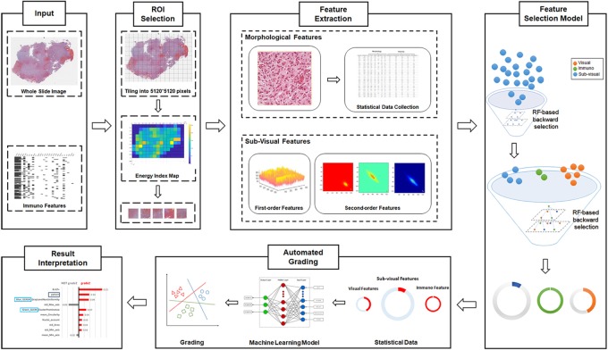
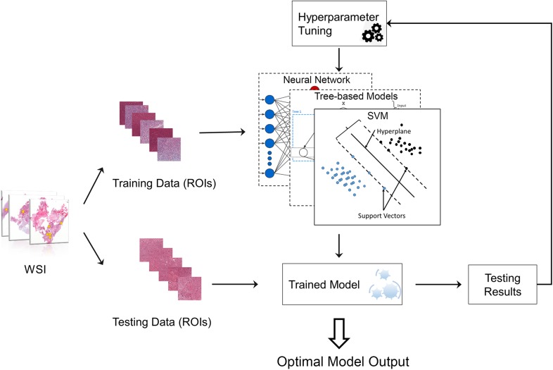
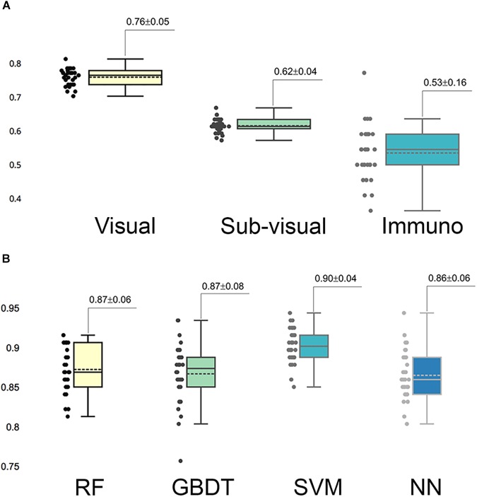
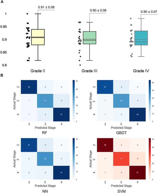
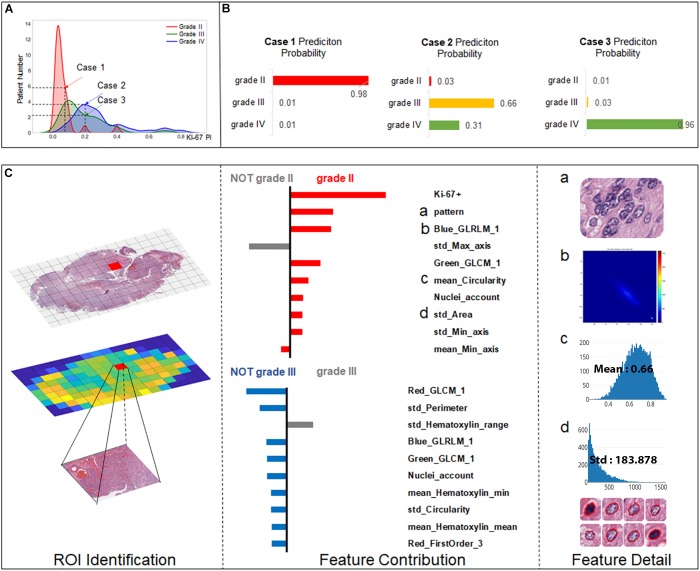
Gliomas are the most common primary malignant brain tumors in adults. Accurate grading is crucial as therapeutic strategies are often disparate for different grades and may influence patient prognosis. This study aims to provide an automated glioma grading platform on the basis of machine learning models. In this paper, we investigate contributions of multi-parameters from multimodal data including imaging parameters or features from the Whole Slide images (WSI) and the proliferation marker Ki-67 for automated brain tumor grading. For each WSI, we extract both visual parameters such as morphology parameters and sub-visual parameters including first-order and second-order features. On the basis of machine learning models, our platform classifies gliomas into grades II, III, and IV. Furthermore, we quantitatively interpret and reveal the important parameters contributing to grading with the Local Interpretable Model-Agnostic Explanations (LIME) algorithm. The quantitative analysis and explanation may assist clinicians to better understand the disease and accordingly to choose optimal treatments for improving clinical outcomes. The performance of our grading model was evaluated with cross-validation, which randomly divided the patients into non-overlapping training and testing sets and repeatedly validated the model on the different testing sets. The primary results indicated that this modular platform approach achieved the highest grading accuracy of 0.90 ± 0.04 with support vector machine (SVM) algorithm, with grading accuracies of 0.91 ± 0.08, 0.90 ± 0.08, and 0.90 ± 0.07 for grade II, III, and IV gliomas, respectively.
神经胶质瘤是成人中最常见的原发性恶性脑肿瘤。准确分级至关重要,因为不同级别的治疗策略往往不同,且可能影响患者预后。本研究旨在基于机器学习模型提供一个自动神经胶质瘤分级平台。在本文中,我们研究了多模态数据中的多参数贡献,包括来自全切片图像(WSI)的成像参数或特征以及增殖标志物Ki-67,用于自动脑肿瘤分级。对于每个WSI,我们提取视觉参数(如形态学参数)和亚视觉参数(包括一阶和二阶特征)。基于机器学习模型,我们的平台将神经胶质瘤分为II级、III级和IV级。此外,我们使用局部可解释模型无关解释(LIME)算法对有助于分级的重要参数进行定量解释和揭示。定量分析和解释可帮助临床医生更好地理解疾病,并据此选择最佳治疗方案以改善临床结果。我们的分级模型性能通过交叉验证进行评估,交叉验证将患者随机分为不重叠的训练集和测试集,并在不同测试集上反复验证模型。主要结果表明,这种模块化平台方法在支持向量机(SVM)算法下实现了最高分级准确率0.90±0.04,II级、III级和IV级神经胶质瘤的分级准确率分别为0.91±0.08、0.90±0.08和0.90±0.07。