Department of Radiology, Faculty of Medicine, University of Peradeniya, Peradeniya, Sri Lanka.
Department of Radiography and Radiotherapy, University of Peradeniya, Peradeniya, Sri Lanka.
Biomed Eng Online. 2022 Aug 1;21(1):52. doi: 10.1186/s12938-022-01022-6.
Diffusion-weighted (DW) imaging is a well-recognized magnetic resonance imaging (MRI) technique that is being routinely used in brain examinations in modern clinical radiology practices. This study focuses on extracting demographic and texture features from MRI Apparent Diffusion Coefficient (ADC) images of human brain tumors, identifying the distribution patterns of each feature and applying Machine Learning (ML) techniques to differentiate malignant from benign brain tumors.
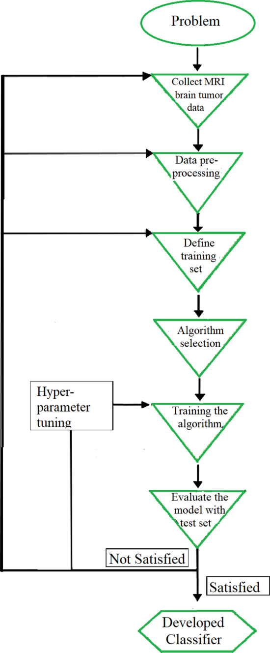
This prospective study was carried out using 1599 labeled MRI brain ADC image slices, 995 malignant, 604 benign from 195 patients who were radiologically diagnosed and histopathologically confirmed as brain tumor patients. The demographics, mean pixel values, skewness, kurtosis, features of Grey Level Co-occurrence Matrix (GLCM), mean, variance, energy, entropy, contrast, homogeneity, correlation, prominence and shade, were extracted from MRI ADC images of each patient. At the feature selection phase, the validity of the extracted features were measured using ANOVA f-test. Then, these features were used as input to several Machine Learning classification algorithms and the respective models were assessed.
According to the results of ANOVA f-test feature selection process, two attributes: skewness (3.34) and GLCM homogeneity (3.45) scored the lowest ANOVA f-test scores. Therefore, both features were excluded in continuation of the experiment. From the different tested ML algorithms, the Random Forest classifier was chosen to build the final ML model, since it presented the highest accuracy. The final model was able to predict malignant and benign neoplasms with an 90.41% accuracy after the hyper parameter tuning process.
This study concludes that the above mentioned features (except skewness and GLCM homogeneity) are informative to identify and differentiate malignant from benign brain tumors. Moreover, they enable the development of a high-performance ML model that has the ability to assist in the decision-making steps of brain tumor diagnosis process, prior to attempting invasive diagnostic procedures, such as brain biopsies.
弥散加权(DW)成像作为一种成熟的磁共振成像(MRI)技术,目前已广泛应用于现代临床放射学中的脑检查。本研究旨在从人脑肿瘤的 MRI 表观扩散系数(ADC)图像中提取人口统计学和纹理特征,确定每个特征的分布模式,并应用机器学习(ML)技术将良恶性脑肿瘤区分开来。
本前瞻性研究使用了 1599 个标注的 MRI 脑 ADC 图像切片,其中 995 个为恶性肿瘤,604 个为良性肿瘤,来自 195 名经影像学诊断和组织病理学证实为脑肿瘤患者。从每位患者的 MRI ADC 图像中提取了人口统计学、平均像素值、偏度、峰度、灰度共生矩阵(GLCM)特征、均值、方差、能量、熵、对比度、同质性、相关性、显著性和阴影。在特征选择阶段,使用 ANOVA f 检验测量提取特征的有效性。然后,将这些特征作为输入应用于几种机器学习分类算法,并评估相应的模型。
根据 ANOVA f 检验特征选择过程的结果,两个属性:偏度(3.34)和 GLCM 同质性(3.45)的 ANOVA f 检验得分最低。因此,在继续进行实验之前,将这两个特征都排除在外。在不同测试的 ML 算法中,选择随机森林分类器来构建最终的 ML 模型,因为它具有最高的准确性。经过超参数调整过程后,最终模型能够以 90.41%的准确率预测良恶性肿瘤。
本研究得出结论,除偏度和 GLCM 同质性外,上述特征可用于识别和区分良恶性脑肿瘤。此外,它们能够开发出高性能的 ML 模型,该模型具有辅助脑肿瘤诊断过程决策步骤的能力,而无需进行侵入性诊断程序,如脑活检。