Ekinci Meliha, Alencar Luciana Magalhães Rebelo, Lopes André Moreni, Santos-Oliveira Ralph, İlem-Özdemir Derya
Faculty of Pharmacy, Department of Radiopharmacy, Ege University, Bornova, Izmir 35040, Turkey.
Biophysics and Nanosystems Laboratory, Department of Physics, Federal University of Maranhão, São Luis 65065-690, Brazil.
J Funct Biomater. 2023 Sep 18;14(9):477. doi: 10.3390/jfb14090477.
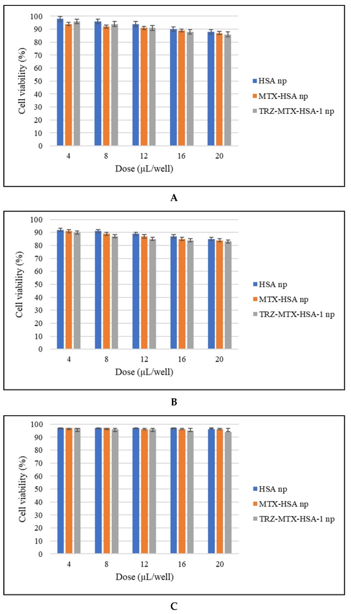
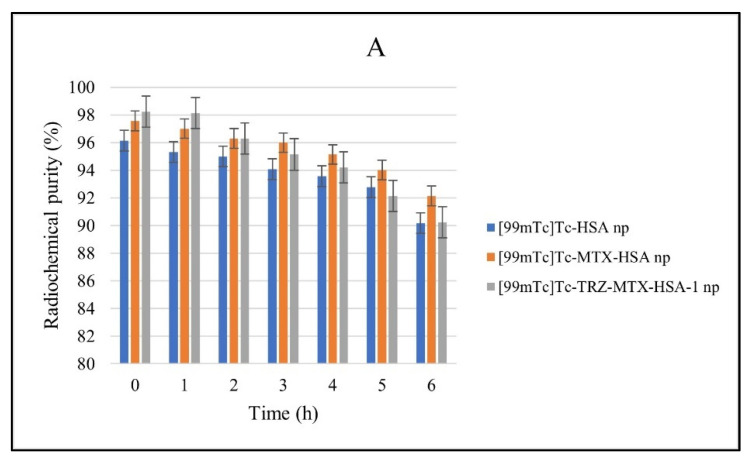
Breast cancer is a leading cause of cancer-related mortality among women worldwide, with millions of new cases diagnosed yearly. Addressing the burden of breast cancer mortality requires a comprehensive approach involving early detection, accurate diagnosis, effective treatment, and equitable access to healthcare services. In this direction, nano-radiopharmaceuticals have shown potential for enhancing breast cancer diagnosis by combining the benefits of nanoparticles and radiopharmaceutical agents. These nanoscale formulations can provide improved imaging capabilities, increased targeting specificity, and enhanced sensitivity for detecting breast cancer lesions. In this study, we developed and evaluated a novel nano-radio radiopharmaceutical, technetium-99m ([Tc]Tc)-labeled trastuzumab (TRZ)-decorated methotrexate (MTX)-loaded human serum albumin (HSA) nanoparticles ([Tc]-TRZ-MTX-HSA), for the diagnosis of breast cancer. In this context, HSA and MTX-HSA nanoparticles were prepared. Conjugation of MTX-HSA nanoparticles with TRZ was performed using adsorption and covalent bonding methods. The prepared formulations were evaluated for particle size, PDI value, zeta (ζ) potential, scanning electron microscopy analysis, encapsulation efficiency, and loading capacity and cytotoxicity on MCF-7, 4T1, and MCF-10A cells. Finally, the nanoparticles were radiolabeled with [Tc]Tc using the direct radiolabeling method, and cellular uptake was performed with the nano-radiopharmaceutical. The results showed the formation of spherical nanoparticles, with a particle size of 224.1 ± 2.46 nm, a PDI value of 0.09 ± 0.07, and a ζ potential value of -16.4 ± 0.53 mV. The encapsulation efficiency of MTX was found to be 32.46 ± 1.12%, and the amount of TRZ was 80.26 ± 1.96%. The labeling with [Tc]Tc showed a high labeling efficiency (>99%). The cytotoxicity studies showed no effect, and the cellular uptake studies showed 97.54 ± 2.16% uptake in MCF-7 cells at the 120th min and were found to have a 3-fold higher uptake in cancer cells than in healthy cells. In conclusion, [Tc]Tc-TRZ-MTX-HSA nanoparticles are promising for diagnosing breast cancer and evaluating the response to treatment in breast cancer patients.
乳腺癌是全球女性癌症相关死亡的主要原因,每年有数百万新病例被诊断出来。应对乳腺癌死亡负担需要采取综合方法,包括早期检测、准确诊断、有效治疗以及公平获得医疗服务。在这方面,纳米放射性药物通过结合纳米颗粒和放射性药物制剂的优势,已显示出增强乳腺癌诊断的潜力。这些纳米级制剂可提供改进的成像能力、更高的靶向特异性以及增强的检测乳腺癌病变的灵敏度。在本研究中,我们开发并评估了一种新型纳米放射性药物,即锝-99m([Tc]Tc)标记的曲妥珠单抗(TRZ)修饰的载有甲氨蝶呤(MTX)的人血清白蛋白(HSA)纳米颗粒([Tc]-TRZ-MTX-HSA),用于乳腺癌的诊断。在此背景下,制备了HSA和MTX-HSA纳米颗粒。使用吸附和共价键合方法将MTX-HSA纳米颗粒与TRZ进行偶联。对制备的制剂进行粒径、多分散指数(PDI)值、zeta(ζ)电位、扫描电子显微镜分析、包封率、载药量以及对MCF-7、4T1和MCF-10A细胞的细胞毒性评估。最后,使用直接放射性标记法用[Tc]Tc对纳米颗粒进行放射性标记,并使用该纳米放射性药物进行细胞摄取实验。结果显示形成了球形纳米颗粒,粒径为224.1±2.46nm,PDI值为0.09±0.07,ζ电位值为-16.4±0.53mV。发现MTX的包封率为32.46±1.12%,TRZ的量为80.26±1.96%。用[Tc]Tc标记显示出高标记效率(>99%)。细胞毒性研究显示无影响,细胞摄取研究显示在第120分钟时MCF-7细胞的摄取率为97.