IRCCS Istituto Tumori Giovanni Paolo II, V.Le O. Flacco, 65, 70124, Bari, Italy.
Department of Basic Medical Sciences Neurosciences and Sense Organs, University of Bari, Piazza G. Cesare, 11, 70124, Bari, Italy.
J Exp Clin Cancer Res. 2023 Sep 28;42(1):251. doi: 10.1186/s13046-023-02808-9.
Clinical drawback in checkpoint inhibitors immunotherapy (ICI) of metastatic melanoma (MM) is monitoring clinical benefit. Soluble forms of PD1(sPD1) and PD-L1(sPD-L1) and extracellular vesicles (EVs) expressing PD1 and PD-L1 have recently emerged as predictive biomarkers of response. As factors released in the blood, EVs and soluble forms could be relevant in monitoring treatment efficacy and adaptive resistance to ICI.
We used pre-therapy plasma samples of 110 MM patients and longitudinal samples of 46 patients. Elisa assay and flow cytometry (FCM) were used to measure sPD-L1 and sPD1 concentrations and the percentage of PD1 EVs and PD-L1 EVs, released from tumor and immune cells in patients subsets. Transwell assays were conducted to investigate the impact of EVs of each patient subset on MM cells invasion and interaction between tumor cells and macrophages or dendritic cells. Viability assays were performed to assess EVs effect on MM cells and organoids sensitivity to anti-PD1. FCM was used to investigate immunosuppressive markers in EVs and immune cells.
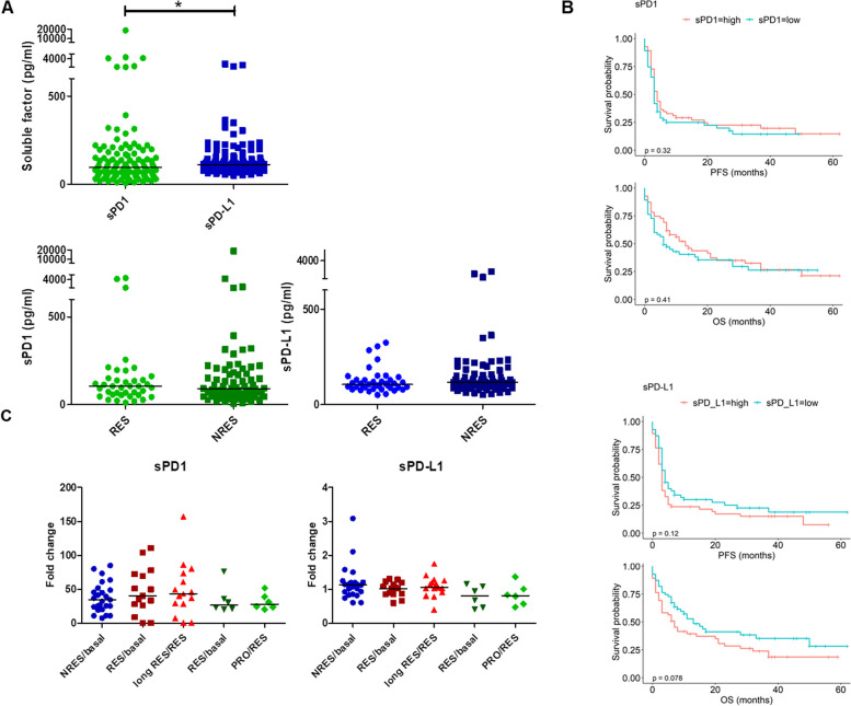
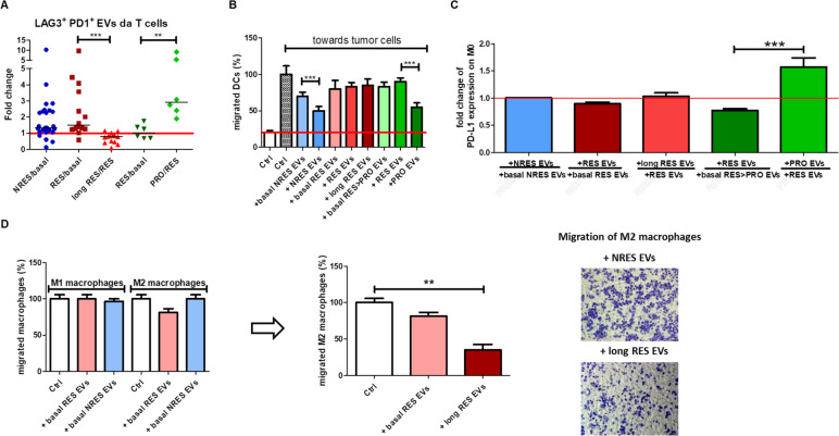
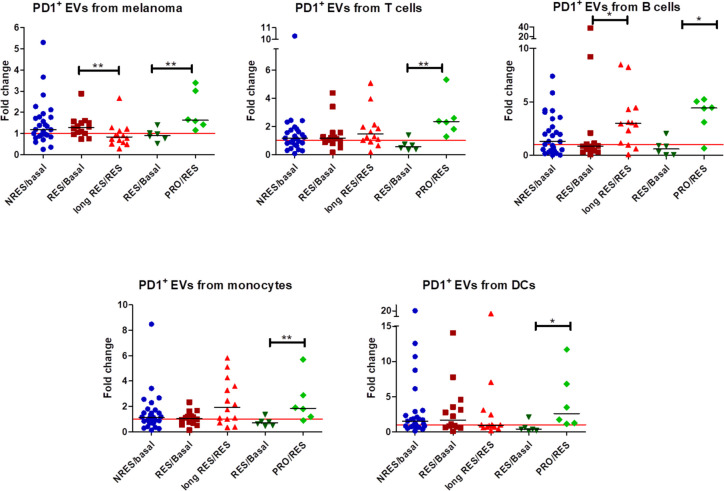
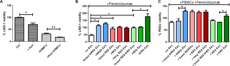
The concentrations of sPD1 and sPD-L1 in pre-treatment and longitudinal samples did not correlate with anti-PD1 response, instead only tumor-derived PD1 EVs decreased in long responders while increased during disease progression in responders. Notably, we observed reduction of T cell derived EVs expressing LAG3 and PD1 in long responders and their increase in responders experiencing progression. By investigating the impact of EVs on disease progression, we found that those isolated from non-responders and from patients with progression disease accelerated tumor cells invasiveness and migration towards macrophages, while EVs of long responders reduced the metastatic potential of MM cells and neo-angiogenesis. Additionally, the EVs of non-responders and of progression disease patients subset reduced the sensitivity of MM cells and organoids of responder to anti-PD1 and the recruitment of dendritic cells, while the EVs of progression disease subset skewed macrophages to express higher level of PDL-1.
Collectively, we suggest that the detection of tumor-derived PD1 + EVs may represent a useful tool for monitoring the response to anti-PD1 and a role for EVs shed by tumor and immune cells in promoting tumor progression and immune dysfunction.
转移性黑色素瘤(MM)的检查点抑制剂免疫治疗(ICI)的临床缺点是监测临床获益。PD1 的可溶性形式(sPD1)和 PD-L1(sPD-L1)以及表达 PD1 和 PD-L1 的细胞外囊泡(EVs)最近已成为反应的预测生物标志物。作为在血液中释放的因子,EVs 和可溶性形式可能与监测治疗效果和对 ICI 的适应性耐药有关。
我们使用了 110 名 MM 患者的治疗前血浆样本和 46 名患者的纵向样本。使用 Elisa 测定法和流式细胞术(FCM)来测量 sPD-L1 和 sPD1 浓度以及来自患者亚群中肿瘤和免疫细胞的 PD1 EVs 和 PD-L1 EVs 的百分比。Transwell 测定法用于研究每个患者亚群的 EVs 对 MM 细胞侵袭的影响以及肿瘤细胞与巨噬细胞或树突状细胞之间的相互作用。进行活力测定以评估 EVs 对 MM 细胞和类器官对抗 PD1 的敏感性的影响。FCM 用于研究 EVs 和免疫细胞中的免疫抑制标志物。
治疗前和纵向样本中的 sPD1 和 sPD-L1 浓度与抗 PD1 反应没有相关性,而是只有长反应者中的肿瘤来源的 PD1 EVs 减少,而在反应者中疾病进展期间增加。值得注意的是,我们观察到长反应者的 T 细胞衍生的 EVs 表达 LAG3 和 PD1 减少,而在进展的反应者中增加。通过研究 EVs 对疾病进展的影响,我们发现来自非反应者和进展疾病患者的那些 EVs加速了肿瘤细胞向巨噬细胞的侵袭和迁移,而长反应者的 EVs 降低了 MM 细胞的转移潜能和新血管生成。此外,非反应者和进展疾病患者亚群的 EVs 降低了 MM 细胞和反应者类器官对抗 PD1 的敏感性以及树突状细胞的募集,而进展疾病亚群的 EVs 使巨噬细胞偏向表达更高水平的 PDL-1。
总之,我们认为检测肿瘤来源的 PD1+EVs 可能是监测抗 PD1 反应的有用工具,并且肿瘤和免疫细胞释放的 EVs 在促进肿瘤进展和免疫功能障碍方面发挥作用。