Discovery Center for Musculoskeletal Recovery, Schoen Adams Research Institute at Spaulding, Charlestown, Massachusetts 02129, United States.
Department of Physical Medicine & Rehabilitation, Harvard Medical School, Boston, Massachusetts 02115, United States.
ACS Nano. 2023 Oct 24;17(20):19640-19651. doi: 10.1021/acsnano.3c02269. Epub 2023 Oct 5.
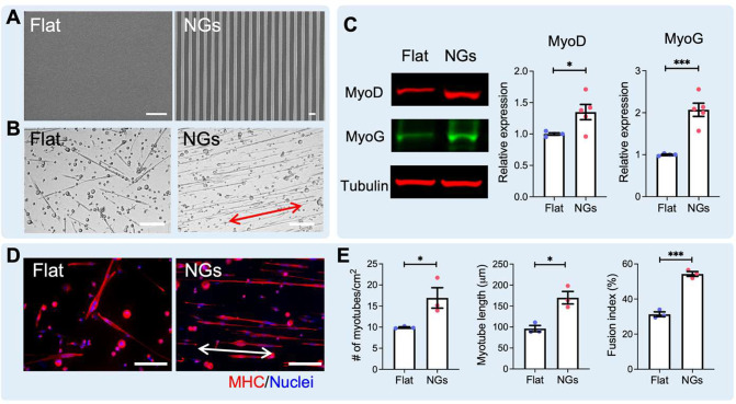
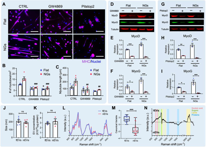
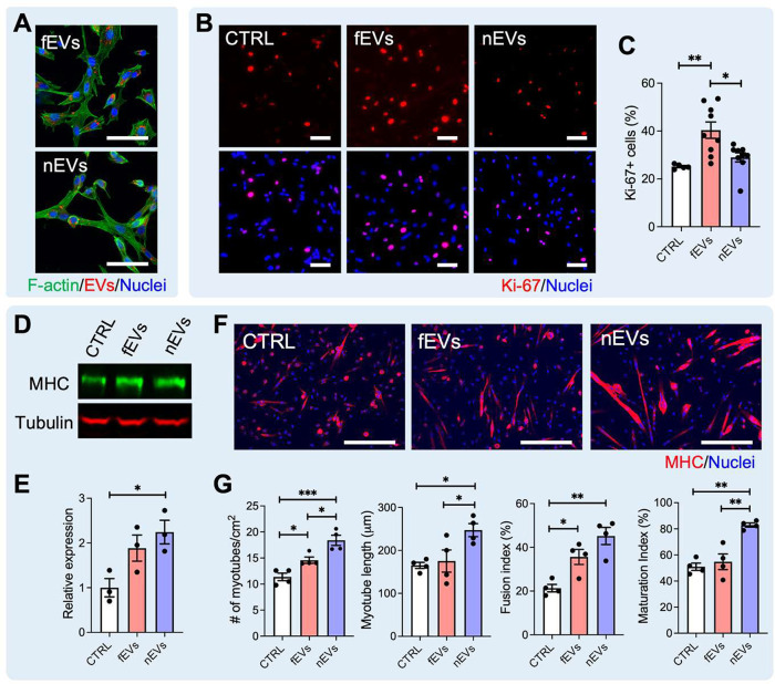
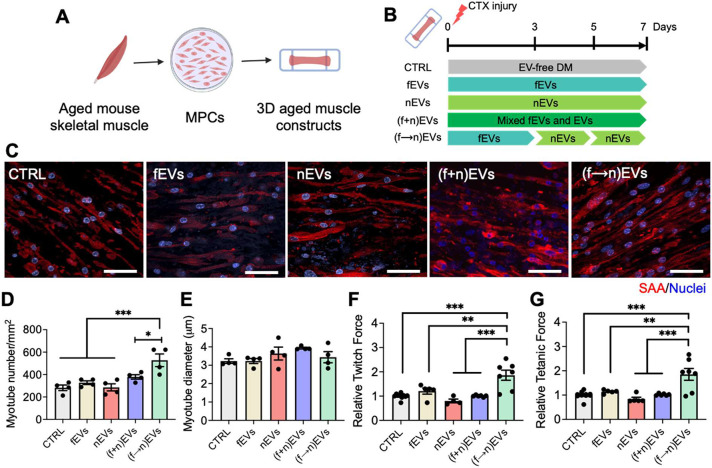
Skeletal muscle regeneration relies on the tightly temporally regulated lineage progression of muscle stem/progenitor cells (MPCs) from activation to proliferation and, finally, differentiation. However, with aging, MPC lineage progression is disrupted and delayed, ultimately causing impaired muscle regeneration. Extracellular vesicles (EVs) have attracted broad attention as next-generation therapeutics for promoting tissue regeneration. As a next step toward clinical translation, strategies to manipulate EV effects on downstream cellular targets are needed. Here, we developed an engineering strategy to tune the therapeutic potential of EVs using nanotopographical cues. We found that EVs released by young MPCs cultured on flat substrates (fEVs) promoted the proliferation of aged MPCs while EVs released by MPCs cultured on nanogratings (nEVs) promoted myogenic differentiation. We then employed a bioengineered 3D muscle aging model to optimize the administration protocol and test the therapeutic potential of fEVs and nEVs in a high-throughput manner. We found that the sequential administration first of fEVs during the phase of MPC proliferative expansion (i.e., 1 day after injury) followed by nEV administration at the stage of MPC differentiation (i.e., 3 days after injury) enhanced aged muscle regeneration to a significantly greater extent than fEVs and nEVs delivered either in isolation or mixed. The beneficial effects of the sequential EV treatment strategy were further validated , as evidenced by increased myofiber size and improved functional recovery. Collectively, our study demonstrates the ability of topographical cues to tune EV therapeutic potential and highlights the importance of optimizing the EV administration strategy to accelerate aged skeletal muscle regeneration.
骨骼肌再生依赖于肌肉干细胞/祖细胞(MPC)从激活到增殖,最终分化的严格时间调控的谱系进展。然而,随着年龄的增长,MPC 谱系进展被打乱和延迟,最终导致肌肉再生受损。细胞外囊泡(EVs)作为促进组织再生的下一代治疗药物引起了广泛关注。作为向临床转化的下一步,需要制定操纵 EV 对下游细胞靶标影响的策略。在这里,我们开发了一种使用纳米形貌线索来调整 EV 治疗潜力的工程策略。我们发现,在平基底上培养的年轻 MPC 释放的 EV(fEVs)促进了衰老 MPC 的增殖,而在纳米光栅上培养的 MPC 释放的 EV(nEVs)促进了成肌分化。然后,我们使用生物工程化的 3D 肌肉衰老模型以高通量的方式优化给药方案并测试 fEVs 和 nEVs 的治疗潜力。我们发现,在 MPC 增殖扩展阶段(即损伤后 1 天)首先给予 fEVs,然后在 MPC 分化阶段(即损伤后 3 天)给予 nEVs 的序贯给药方案,比单独或混合给予 fEVs 和 nEVs 更能显著增强衰老肌肉的再生。序贯 EV 治疗策略的有益效果进一步得到验证,表现为肌纤维大小增加和功能恢复改善。总之,我们的研究表明,形貌线索能够调整 EV 的治疗潜力,并强调了优化 EV 给药策略以加速衰老骨骼肌再生的重要性。